Heart Diagram Labeled: Chambers, Valves & Coronary Arteries
Heart diagram labeled: the four chambers, heart valves, coronary arteries (LAD, RCA, circumflex), the SA node and conduction system, and heart attacks.
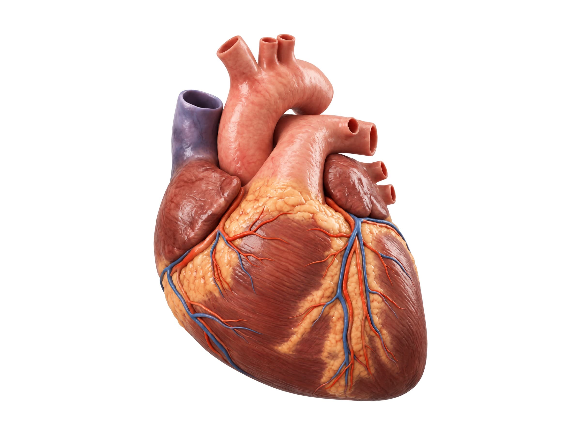
The heart is a muscular pump about the size of a fist that sits in the middle of the chest, between the lungs and behind the breastbone, with its tip (the apex) pointing down and to the left. Heart anatomy comes down to four chambers (two atria and two ventricles), four valves that keep blood moving in one direction, the great vessels that carry blood in and out, the coronary arteries that supply the heart muscle itself, and an electrical conduction system that sets the heartbeat. Knowing how these parts fit together is the first step to reading an echocardiogram or angiogram report, explaining a heart attack to a patient, or illustrating a cardiac injury or malpractice claim.
Diagram of the heart: parts of the heart labeled
The hero illustration above shows the heart from the front. Because the heart is rotated in the chest, most of the front surface is the right ventricle; the left ventricle forms the left border and the apex, and the right atrium forms the right border. The aorta rises from the center and arches over to the left, giving off the brachiocephalic trunk, left common carotid artery and left subclavian artery; the pulmonary trunk rises in front of it and divides under the arch.
- Atria: the two thin-walled upper chambers that receive blood. Each has a small ear-shaped pouch, the right auricle and left auricle.
- Ventricles: the two thick-walled lower chambers that pump blood out of the heart.
- Great vessels: the superior vena cava and inferior vena cava bring blood from the body to the right atrium; the pulmonary artery carries it to the lungs; the pulmonary veins bring it back to the left atrium; and the aorta carries it to the body.
- Coronary arteries: the vessels on the surface of the heart that supply the heart muscle.
Where the heart sits in the chest
The heart lies in the mediastinum, the central compartment of the chest between the two lungs. About two-thirds of it lies to the left of the midline. Above it, the aortic arch curves over the left main bronchus, the superior vena cava runs down on the right, and the trachea lies behind the great vessels.
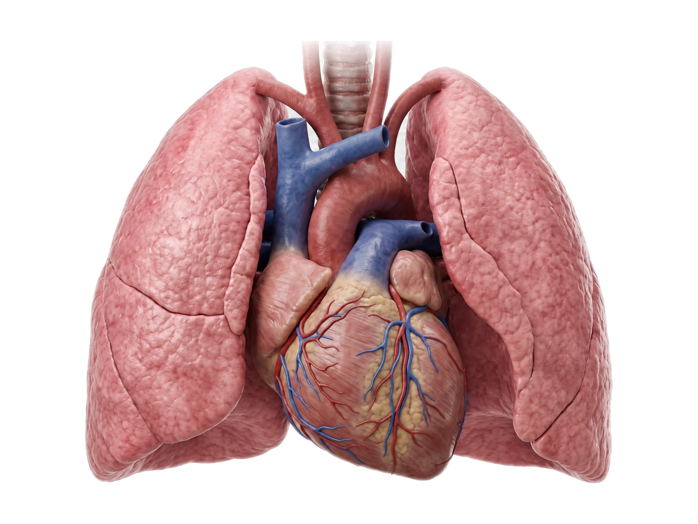
The heart and the roots of the great vessels are wrapped in the pericardium, a two-layered sac. The outer fibrous pericardium is tough and anchors the heart to the diaphragm and breastbone; the inner serous pericardium lines the sac and covers the heart surface, with a thin film of fluid between the layers so the heart can beat without friction.
Chambers of the heart
The heart has four chambers: two thin-walled atria on top that receive blood, and two thick-walled ventricles below that pump it out. A wall called the septum divides the right side from the left, so oxygen-poor and oxygen-rich blood never mix after birth: the interatrial septum separates the atria and the much thicker interventricular septum separates the ventricles. The cross section below, cut from front to back through the whole heart, shows all four chambers and all four valves at once.
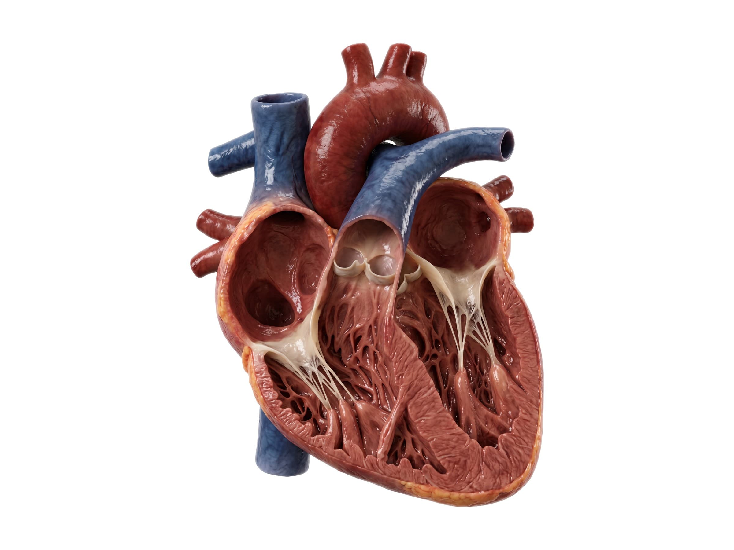
- Right atrium: receives blood from the body through the superior and inferior vena cava.
- Right ventricle: pumps blood through the pulmonary valve to the lungs. It wraps around the front of the left ventricle and is crescent-shaped in cross section.
- Left atrium: receives oxygen-rich blood from the lungs through the four pulmonary veins.
- Left ventricle: the main pumping chamber, with a wall about three times thicker than the right ventricle's, pumping blood through the aortic valve to the whole body.
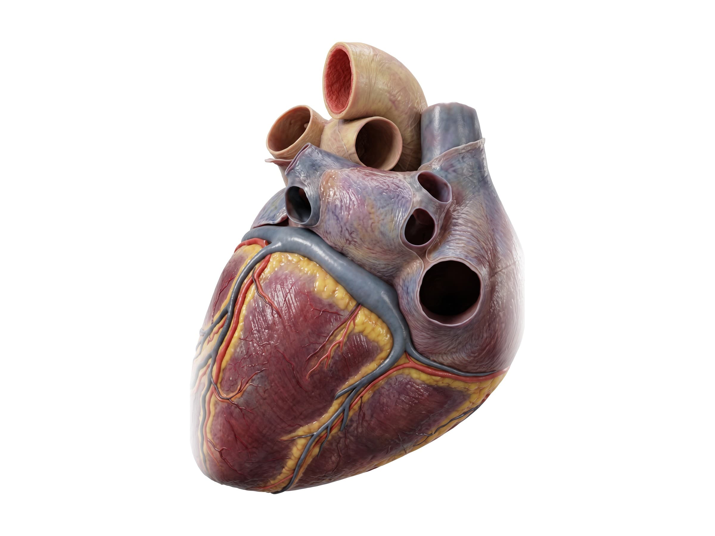
The back of the heart is formed mostly by the left atrium, with the four pulmonary veins entering it. The coronary sinus, the large vein that drains the heart muscle, runs in the groove between the atria and ventricles and empties into the right atrium.
Right atrium and right ventricle
The right atrium receives oxygen-poor blood from the superior and inferior vena cava and from the coronary sinus. Its wall has a smooth back part and a ridged front part lined with pectinate muscles, divided by a ridge called the crista terminalis. On the wall it shares with the left atrium (the interatrial septum) is the fossa ovalis, an oval dip that marks the opening, the foramen ovale, that let blood bypass the lungs before birth.
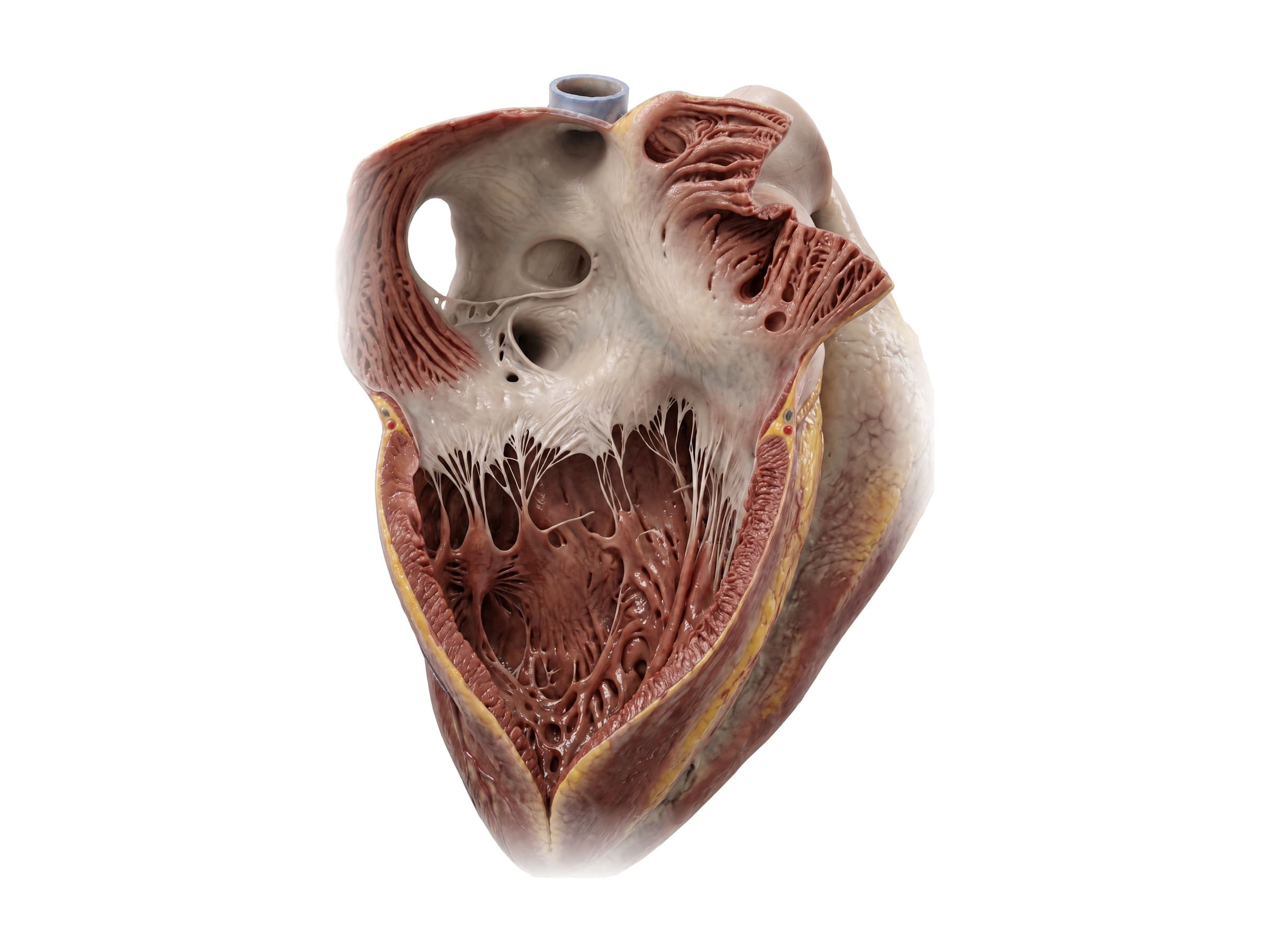
Blood passes through the tricuspid valve into the right ventricle, which pumps it through the pulmonary valve into the lungs. The right ventricle has a thinner wall than the left because it pumps against much lower pressure.
Left atrium and left ventricle
The left atrium receives oxygen-rich blood from the lungs through the four pulmonary veins and passes it through the mitral valve into the left ventricle. The left ventricle's wall is about three times thicker than the right's, and its inner surface is covered with ridges of muscle called trabeculae carneae.
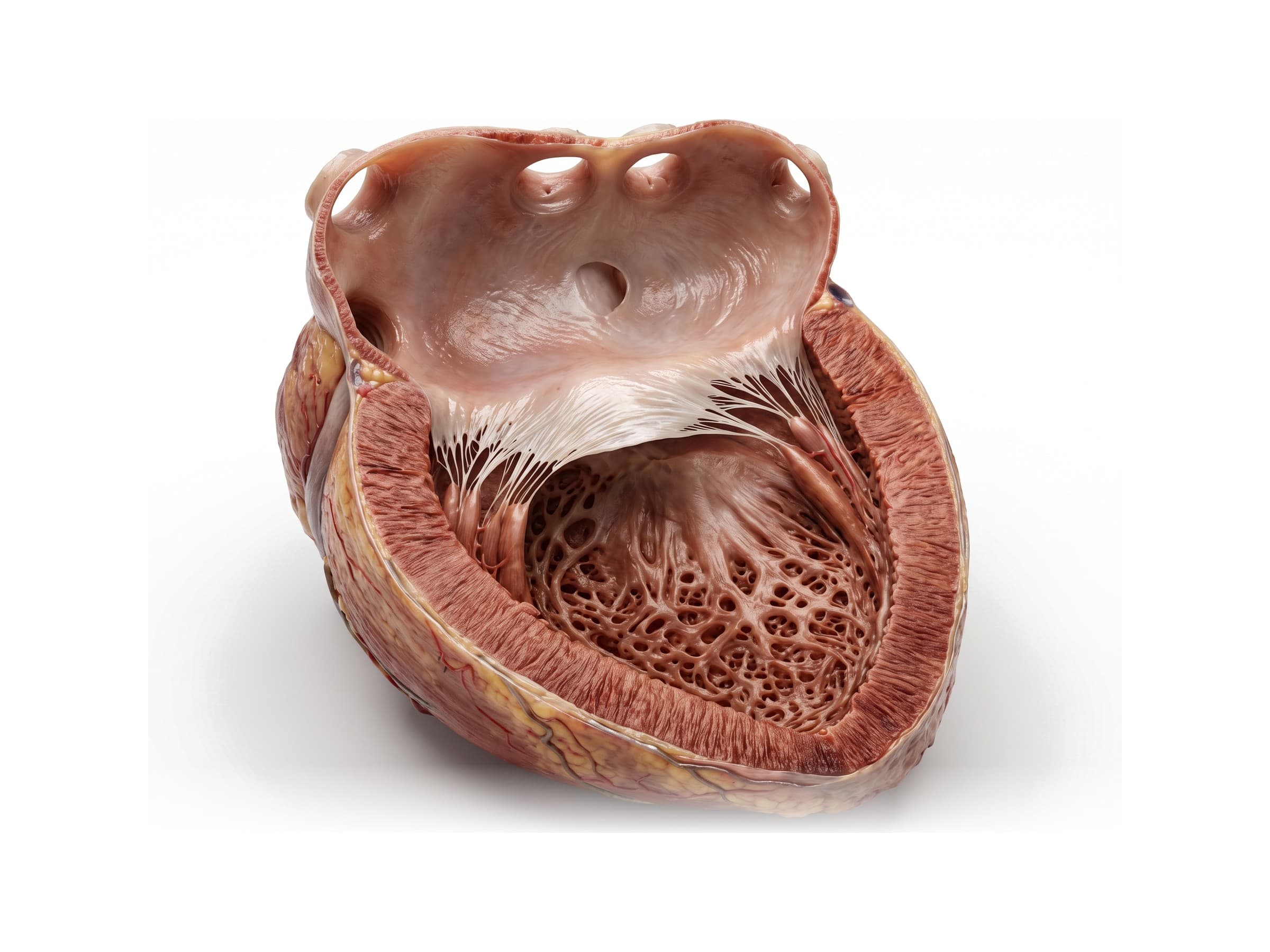
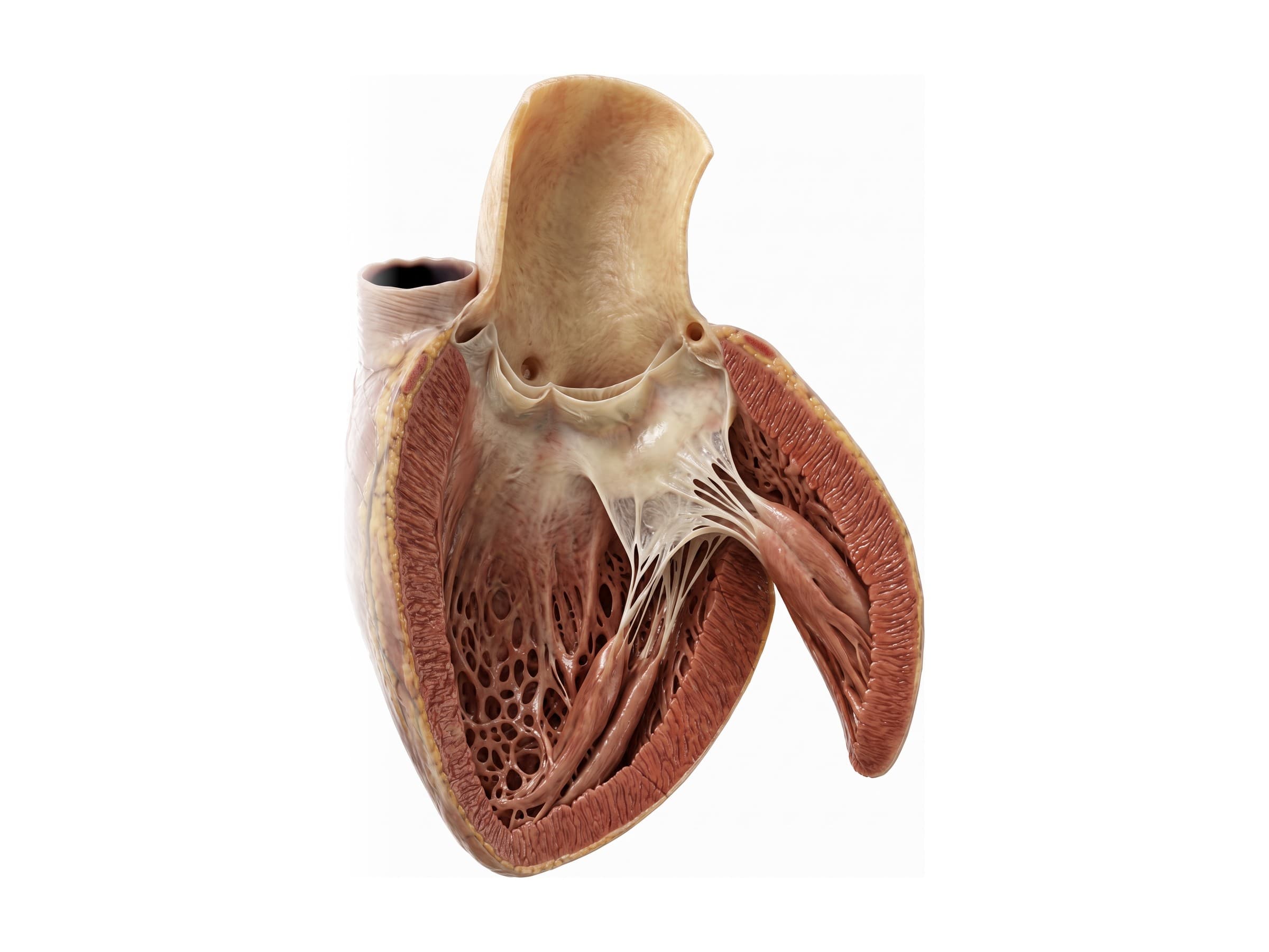
The muscular wall between the two ventricles is the interventricular septum. When the left ventricle contracts, it pushes blood up through the aortic valve into the aorta. The openings of the right and left coronary arteries sit in the wall of the aorta just above the cusps of this valve.
Heart valves
Four valves keep blood flowing forward. Seen from above with the atria removed, they sit together in a ring of dense connective tissue called the fibrous skeleton of the heart.
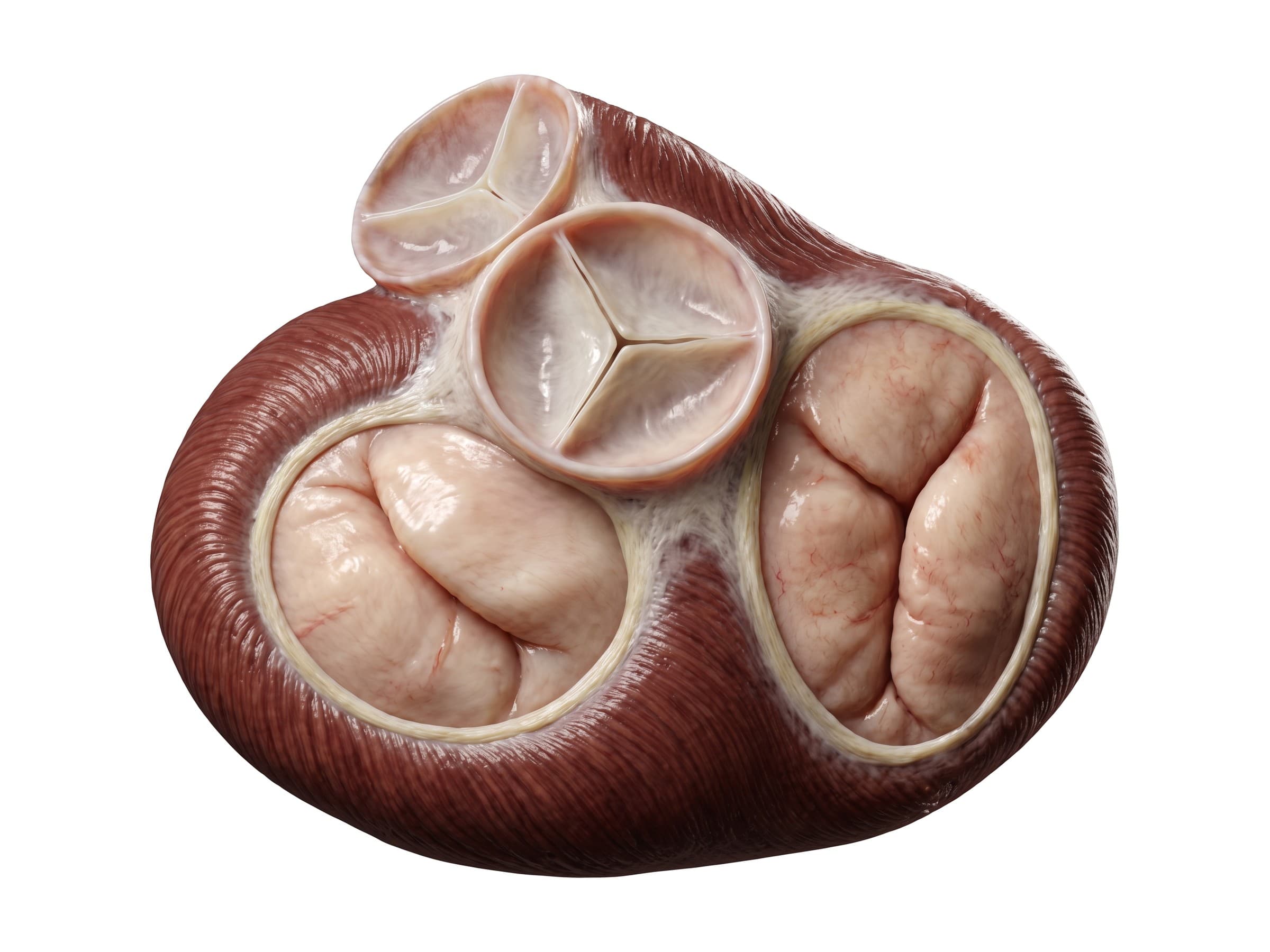
- Tricuspid valve: between the right atrium and right ventricle, with three cusps (anterior, posterior and septal).
- Pulmonary valve: between the right ventricle and the pulmonary artery, with three half-moon (semilunar) cusps.
- Mitral valve (bicuspid valve): between the left atrium and left ventricle, with two cusps, anterior and posterior. It is the valve most often affected by prolapse and regurgitation.
- Aortic valve: between the left ventricle and the aorta, with three semilunar cusps. Narrowing of this valve (aortic stenosis) is the most common valve disease in older adults.
The tricuspid and mitral valves are tied to the ventricle walls by thin cords, the chordae tendineae, which attach to papillary muscles. When the ventricles contract, the papillary muscles tighten the cords so the cusps close without flipping back into the atria. A ruptured papillary muscle after a heart attack causes sudden, severe mitral regurgitation.
Blood flow through the heart
- Oxygen-poor blood from the body enters the right atrium through the superior and inferior vena cava.
- It passes through the tricuspid valve into the right ventricle.
- The right ventricle pumps it through the pulmonary valve into the pulmonary artery and the lungs, where it picks up oxygen.
- Oxygen-rich blood returns through the pulmonary veins to the left atrium.
- It passes through the mitral valve into the left ventricle.
- The left ventricle pumps it through the aortic valve into the aorta and out to the body.
Both sides work at the same time: the two atria contract together, then the two ventricles.
Coronary arteries
The heart muscle cannot use the blood passing through its chambers; it gets its own supply from the coronary arteries. There are two, the left and the right coronary artery, and they are the first branches of the aorta. They run over the surface of the heart in the fat-filled grooves between the chambers, and their branches dive into the muscle. Because the heart muscle is squeezed during each beat, most coronary blood flow happens between beats, while the heart relaxes.
Where the coronary arteries begin
Both coronary arteries open from the wall of the aorta just above the aortic valve, in two of its three bulges, the aortic sinuses (sinuses of Valsalva). The left coronary artery opens above the left coronary cusp, the right coronary artery above the right coronary cusp, and the third cusp, with no artery above it, is the non-coronary cusp. An artery that starts from the wrong sinus (an anomalous coronary artery) can be squeezed between the aorta and pulmonary artery during exercise and is a known cause of sudden death in young athletes.
Branches of the coronary arteries
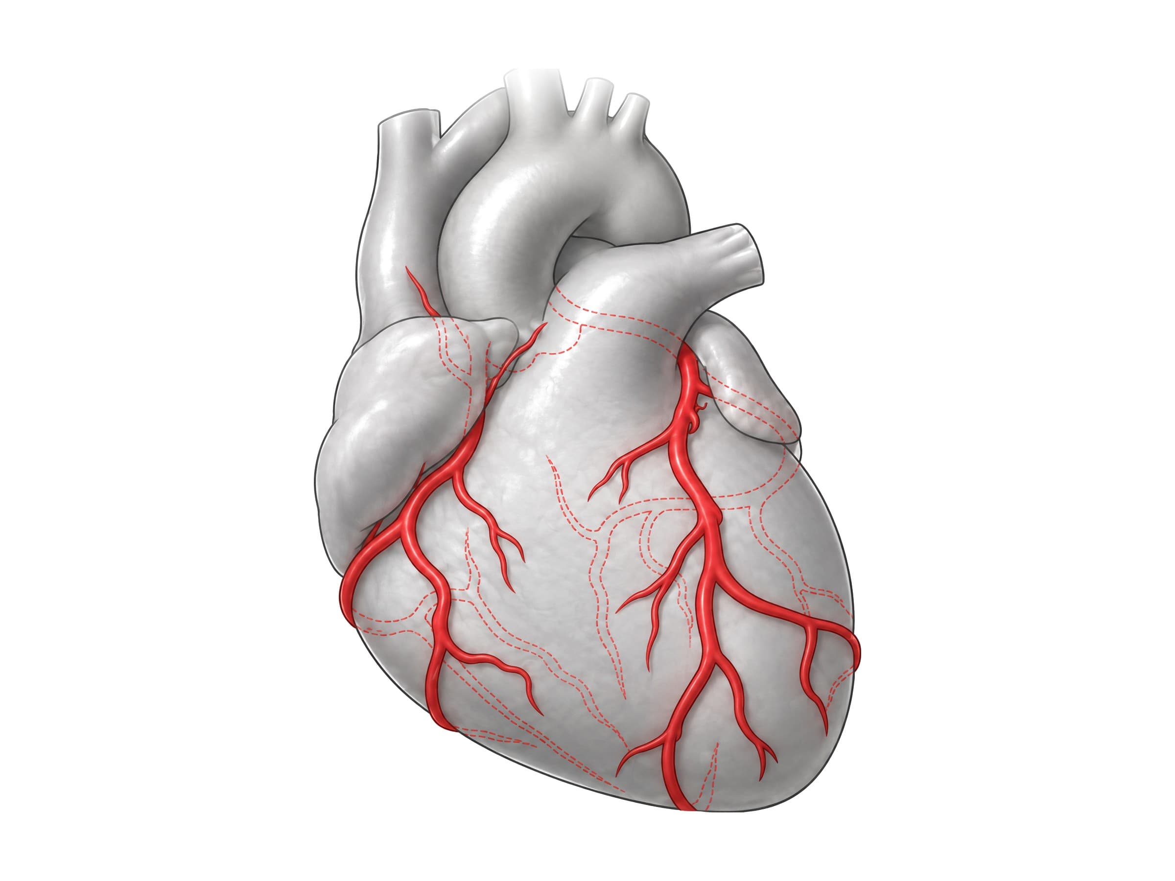
Left coronary artery
- Left main coronary artery: a short trunk, usually 1 to 2 cm long, that passes behind the pulmonary trunk and divides into the LAD and the circumflex. Because it feeds both, a blockage here threatens most of the left ventricle. In roughly 15% to 30% of people it splits into three instead, the middle branch being the ramus intermedius.
- Left anterior descending artery (LAD), also called the anterior interventricular artery: runs down the groove between the ventricles on the front of the heart, often wrapping around the apex onto the underside. It supplies the front wall of the left ventricle, the front two-thirds of the interventricular septum, and the apex: the largest territory of any coronary artery.
- Diagonal branches: run diagonally from the LAD across the front-side wall of the left ventricle.
- Septal perforators: dive straight into the interventricular septum and supply the bundle branches of the conduction system.
- Circumflex artery (left circumflex, LCx): runs to the left in the groove between the left atrium and left ventricle and around to the back of the heart. It supplies the side and back wall of the left ventricle and the left atrium.
- Obtuse marginal branches: run down the rounded left border of the heart (its "obtuse margin") over the side wall of the left ventricle.
Right coronary artery
- Right coronary artery (RCA): runs in the groove between the right atrium and right ventricle, down the right border of the heart and around onto its underside. It supplies the right atrium, the right ventricle, and usually the underside of the left ventricle.
- Conus artery: the first branch, supplying the outflow tract of the right ventricle under the pulmonary valve.
- Sinoatrial (SA) nodal artery: comes from the RCA in about 60% of people (from the circumflex in the rest) and supplies the heart's pacemaker.
- Anterior right ventricular branches: run across the front wall of the right ventricle.
- Acute marginal artery: runs along the sharp lower right border of the heart (its "acute margin").
- AV nodal artery: comes from the RCA where it crosses the back of the heart in most people, supplying the AV node. This is why blockage of the RCA often causes a slow heart rate or heart block.
The great cardiac vein runs alongside the LAD and then the circumflex, collecting blood from the front of the left ventricle, while the small anterior cardiac veins drain the right ventricle directly into the right atrium.
The back of the heart
On the back and underside of the heart, the circumflex and the right coronary artery approach each other in the groove between the atria and ventricles. The posterior descending artery (PDA), also called the posterior interventricular artery, runs down the groove between the ventricles on the underside of the heart and supplies the back third of the interventricular septum and the inferior wall of the left ventricle. The middle cardiac vein runs beside it, and the large coronary sinus, which collects blood from the great, middle and small cardiac veins, empties into the right atrium.
Coronary dominance
Whichever artery gives off the posterior descending artery is called dominant. About 80% to 85% of people are right dominant (the PDA comes from the RCA), about 5% to 10% are left dominant (from the circumflex), and the rest are co-dominant, with branches from both. Dominance matters clinically: in a left-dominant heart, the circumflex also supplies the underside of the left ventricle and the AV node, so a blockage of the left main or circumflex cuts off blood to a much larger part of the heart.
Which artery supplies which part of the heart
| Territory | Usual artery | Heart attack it causes |
|---|---|---|
| Front wall of the left ventricle, apex, front two-thirds of the septum | LAD | Anterior (and anteroseptal) infarct |
| Side (lateral) wall of the left ventricle | Circumflex and diagonal branches | Lateral infarct |
| Underside (inferior) wall of the left ventricle, back third of the septum | RCA, through the posterior descending artery | Inferior infarct |
| Right ventricle | RCA | Right ventricular infarct |
| SA node and AV node | Usually RCA | Slow heart rate, heart block |
On an ECG, these territories map onto groups of leads, which is how a cardiologist can predict the blocked artery before an angiogram: changes in leads V1 to V4 point to the LAD, I, aVL, V5 and V6 to the circumflex, and II, III and aVF to the right coronary artery.
How coronary arteries are imaged
A coronary angiogram threads a thin catheter from the wrist or groin into the opening of each coronary artery and injects dye while taking X-ray video, showing narrowing and blockages and allowing a stent to be placed in the same procedure. CT coronary angiography shows the arteries without a catheter, and a calcium score CT measures the calcified plaque in their walls. Some people have a myocardial bridge, a stretch of an artery, usually the LAD, that dives into the muscle instead of running on its surface and is squeezed with every beat.
Electrical conduction system of the heart: SA node and AV node
Each heartbeat starts in the sinoatrial (SA) node, a small cluster of pacemaker cells in the wall of the right atrium near the superior vena cava. The impulse spreads across both atria, making them contract, and reaches the atrioventricular (AV) node in the floor of the right atrium. The AV node briefly delays the signal so the ventricles can fill, then passes it to the bundle of His, which runs down the interventricular septum and splits into the right and left bundle branches. These end in a network of Purkinje fibers in the ventricle walls, which make both ventricles contract from the apex upward.
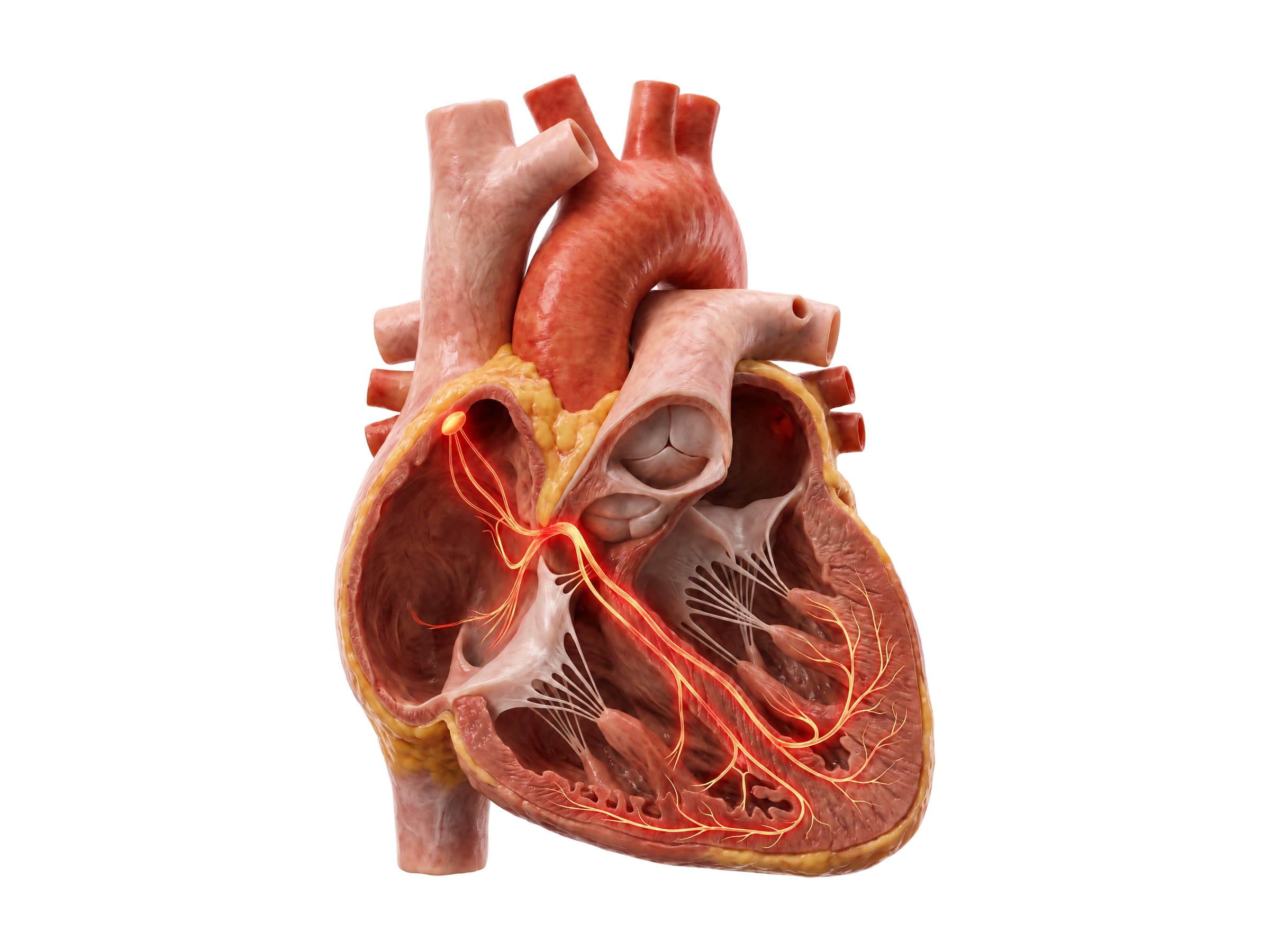
Damage to this pathway causes heart block (a delayed or blocked signal from the atria to the ventricles) or bundle branch blocks, which show up on an electrocardiogram (ECG) and may need a pacemaker.
Layers of the heart wall
- Epicardium: the thin outer layer on the surface of the heart (the inner layer of the serous pericardium), carrying the coronary arteries and fat.
- Myocardium: the thick middle layer of cardiac muscle that does the pumping. It is thickest in the left ventricle.
- Endocardium: the smooth inner lining of the chambers and valves. Infection of this lining and the valves is endocarditis.
Common heart conditions and how they appear on imaging
- Heart attack (myocardial infarction): a coronary artery is blocked, usually by a blood clot on a ruptured cholesterol plaque, and the heart muscle it supplies starts to die. A blockage of the LAD causes an anterior infarct; when the blockage is in the left main or the start of the LAD it is called a widowmaker. Diagnosed by ECG changes and a rise in troponin in the blood; coronary angiography shows the blocked artery, and echocardiography shows the damaged wall not moving normally.
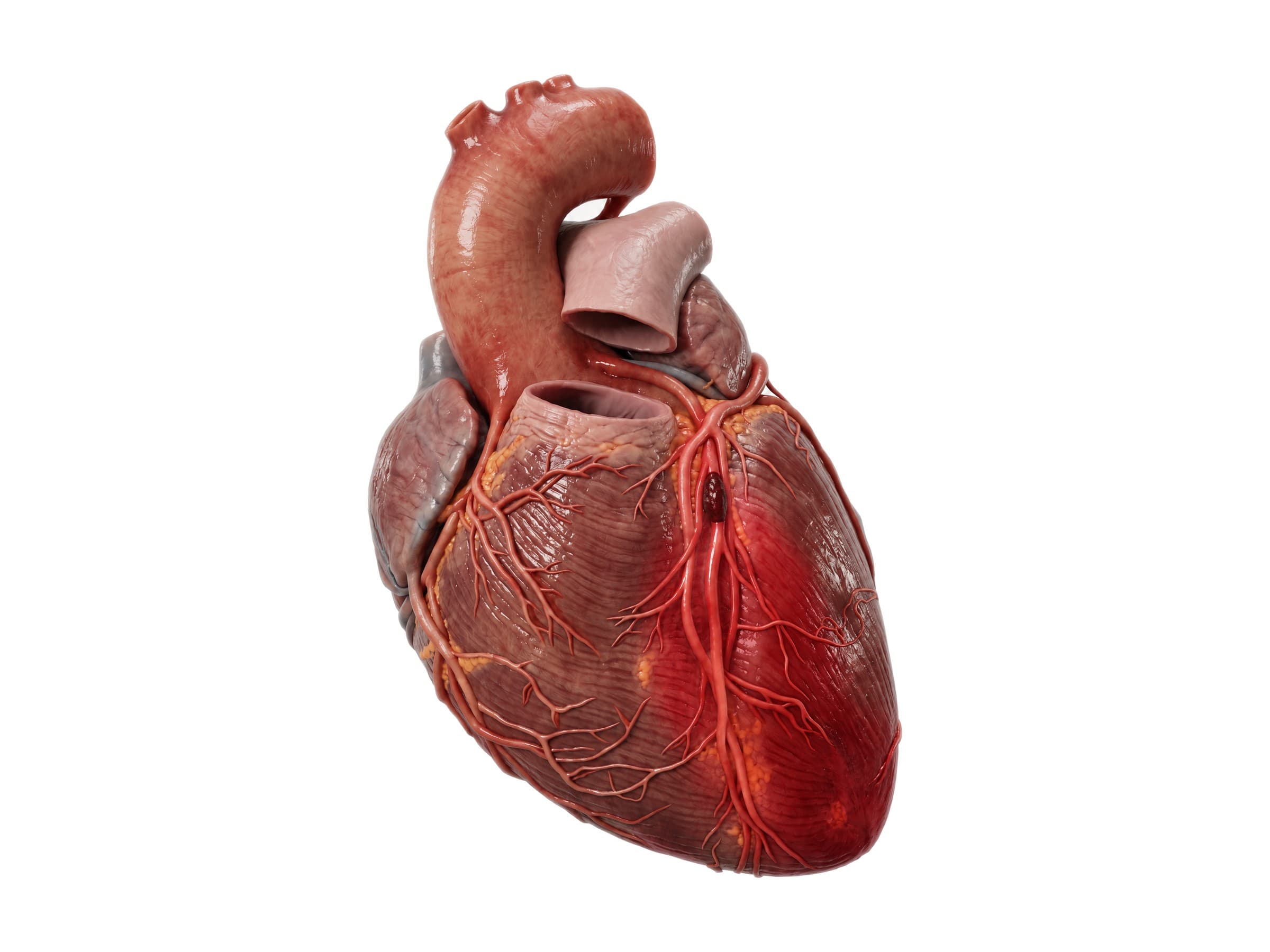
- Coronary artery disease: narrowing of the coronary arteries by plaque, causing chest pain on exertion (angina). Seen on CT coronary angiography or invasive angiography, and treated with stents or bypass surgery.
- Valve disease: narrowing (stenosis) or leaking (regurgitation) of a valve, most often the aortic and mitral valves. Echocardiography shows the valve motion and the flow through it.
- Aortic dissection: a tear in the inner wall of the aorta that lets blood split its layers apart. A surgical emergency, diagnosed with CT angiography.
- Cardiac tamponade: fluid or blood collecting in the pericardial sac under pressure and squeezing the heart, so it cannot fill. Seen on echocardiography; it can follow penetrating chest trauma, a ruptured heart wall, or a complication of a cardiac procedure.
- Blunt cardiac injury: bruising of the heart muscle (myocardial contusion) from a steering-wheel impact or a fall onto the chest, usually affecting the right ventricle because it lies directly behind the breastbone. Suspected from ECG changes and troponin, and evaluated with echocardiography.
- Arrhythmias: abnormal heart rhythms from problems in the conduction system, such as atrial fibrillation or heart block, diagnosed by ECG.
How the heart is shown in a medical malpractice or personal injury case
A labeled illustration of normal heart anatomy is the baseline exhibit in cardiac claims. In medical malpractice cases, the most common are a missed or delayed heart attack diagnosis, a complication of a stent, catheter or valve procedure, and a missed aortic dissection. The exhibit shows a jury where the coronary arteries run and which part of the heart each one supplies, before the case-specific exhibit is introduced: the blocked artery, the area of damaged muscle, and the timeline of symptoms, ECGs and troponin results against the time it took to open the artery. In personal injury claims, the same anatomy explains blunt cardiac injury and tamponade after a motor vehicle collision, tying the injury to the mechanism of injury.
Natomy creates custom medical legal illustrations of cardiac injuries and procedures for demand packages and trial exhibits, and the role of these visuals in court is covered in demonstrative evidence. For more on how the heart's structure is drawn, see the 3D anatomy of the heart and cardiac muscle drawing guides. For the spine and ribs behind the heart, see thoracic spine anatomy, and for the brain and its blood supply, brain anatomy.
FAQ
What are the 4 chambers of the heart?+
The heart has two upper chambers, the right and left atria, and two lower chambers, the right and left ventricles. The right atrium receives oxygen-poor blood from the body and passes it to the right ventricle, which pumps it to the lungs. The left atrium receives oxygen-rich blood from the lungs and passes it to the left ventricle, which pumps it out to the whole body through the aorta. The left ventricle has the thickest wall because it does the most work.
What are the 4 valves of the heart?+
The tricuspid valve sits between the right atrium and right ventricle, and the pulmonary valve between the right ventricle and the pulmonary artery. On the left side, the mitral valve sits between the left atrium and left ventricle, and the aortic valve between the left ventricle and the aorta. The tricuspid and mitral valves are anchored by chordae tendineae and papillary muscles; the pulmonary and aortic valves have three half-moon-shaped cusps each.
What is the order of blood flow through the heart?+
Oxygen-poor blood enters the right atrium from the superior and inferior vena cava, passes through the tricuspid valve into the right ventricle, then through the pulmonary valve into the pulmonary artery and the lungs. Oxygen-rich blood returns through the pulmonary veins to the left atrium, passes through the mitral valve into the left ventricle, and leaves through the aortic valve into the aorta and the rest of the body.
What are the main coronary arteries?+
The two coronary arteries arise from the aorta just above the aortic valve. The left main coronary artery divides into the left anterior descending artery (LAD), which runs down the front of the heart, and the circumflex artery, which wraps around the left side. The right coronary artery runs around the right side of the heart and usually gives off the posterior descending artery on the underside. Together they supply the heart muscle itself with blood.
What is coronary dominance?+
Coronary dominance describes which artery gives off the posterior descending artery, which supplies the underside of the heart and the back of the septum. About 80% to 85% of people are right dominant (the posterior descending artery comes from the right coronary artery), about 5% to 10% are left dominant (it comes from the circumflex), and the rest are co-dominant. In a left-dominant heart, the left coronary artery supplies almost the entire left ventricle, so a blockage there is more dangerous.
Why is it called the widowmaker heart attack?+
A widowmaker is a heart attack caused by a blockage of the left main coronary artery or the very beginning of the left anterior descending artery (LAD). The LAD supplies the front wall of the left ventricle and most of the septum, a large share of the heart's pumping muscle, so a blockage high up can cause sudden, extensive damage and a high risk of cardiac arrest if blood flow is not restored quickly.
Where is the SA node?+
The sinoatrial (SA) node is a small cluster of specialized cells in the wall of the right atrium, near where the superior vena cava enters. It is the heart's natural pacemaker: it fires about 60 to 100 times a minute at rest, and each impulse spreads across both atria, makes them contract, and then reaches the atrioventricular (AV) node, which relays it to the ventricles through the bundle of His and the Purkinje fibers.
What is the pericardium?+
The pericardium is the two-layered sac that surrounds the heart. Its tough outer fibrous layer anchors the heart in the chest, and its thin inner serous layer lines the sac and covers the heart surface, with a small amount of lubricating fluid between them. Inflammation of this sac is pericarditis, and fluid or blood collecting inside it under pressure can squeeze the heart, a condition called cardiac tamponade.
What is the difference between the myocardium, endocardium and epicardium?+
They are the three layers of the heart wall. The epicardium is the thin outer layer on the surface of the heart, which carries the coronary arteries and fat. The myocardium is the thick middle layer of cardiac muscle that does the pumping. The endocardium is the smooth inner lining of the chambers and valves, in direct contact with the blood.
Need a custom illustration of this joint?
Generate a labeled or exhibit-ready figure for a specific patient, injury, or case in minutes.
Try Natomy →