Abdominal Organs Diagram: Stomach, Liver, Kidneys & Bowel Labeled
Labeled abdominal organs diagram: stomach, liver, gallbladder, pancreas, spleen, kidneys, small intestine and colon, plus quadrants and common injuries.
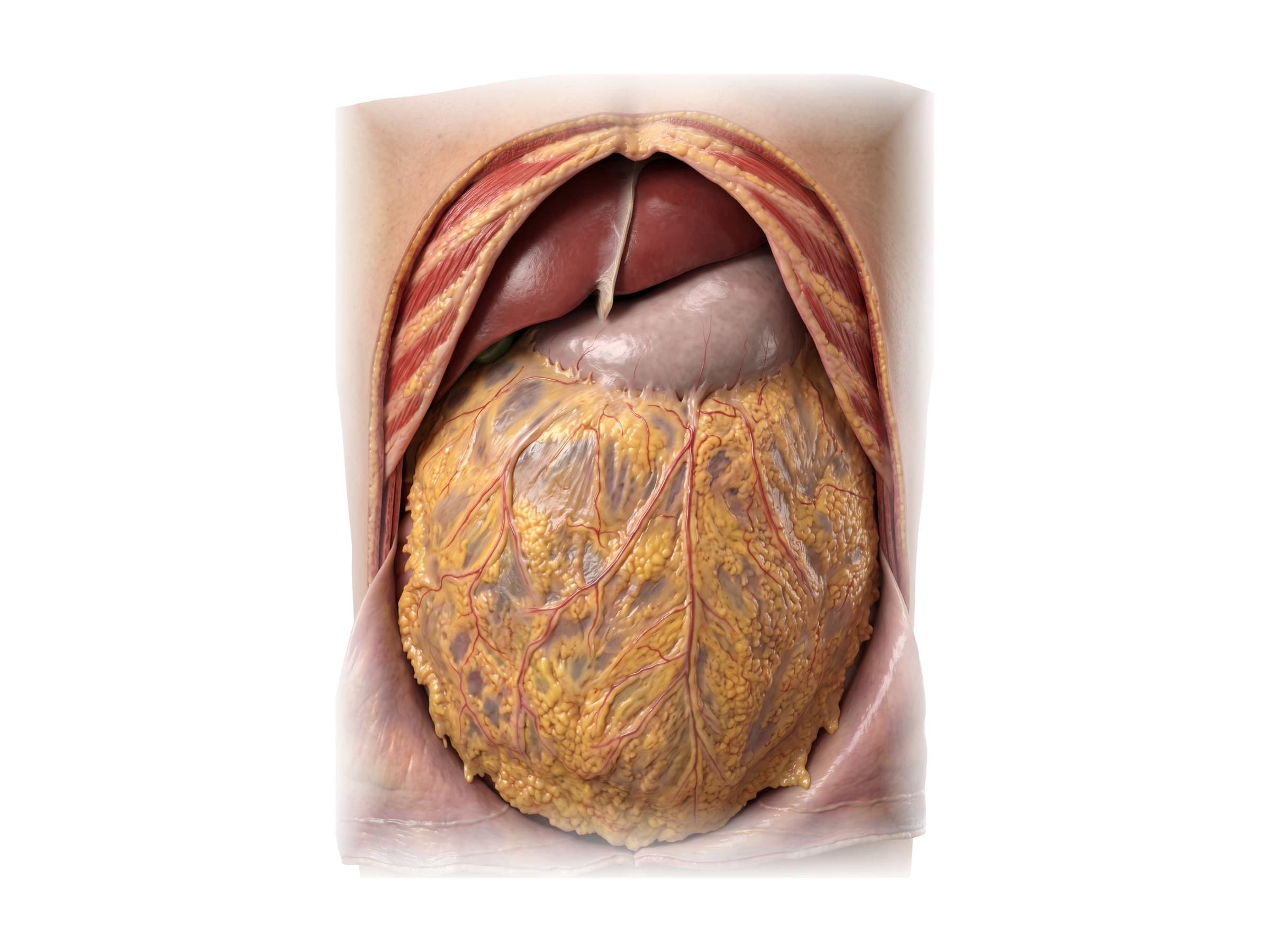
The abdominal organs are the organs that sit in the abdominal cavity, the space between the diaphragm above and the pelvis below. They include the stomach, the small intestine (duodenum, jejunum and ileum), the large intestine (cecum, appendix and colon), the liver, the gallbladder, the pancreas and the spleen, along with the kidneys, adrenal glands and ureters, which lie behind the abdominal lining against the back wall. The digestive organs are wrapped in a thin lining called the peritoneum and covered at the front by a fatty apron, the greater omentum. The labeled diagrams on this page show the abdominal organs layer by layer, from the front view in place, through the stomach, liver, pancreas and bowel, down to the kidneys on the back wall, for reading CT and operative reports, explaining a diagnosis or operation to a patient, and illustrating an injury or malpractice claim.
Abdominal organs diagram: front view
Opening the abdominal wall, as in the image at the top of this page, shows only a few organs at first. The liver fills the upper right under the ribs and crosses the midline, divided into a larger right lobe and a smaller left lobe by the falciform ligament. The tip of the gallbladder peeks out under the lower edge of the liver on the right. The stomach sits under the ribs on the left. Everything below is hidden by the greater omentum, a fatty sheet that hangs from the lower border of the stomach over the intestines. The omentum can move toward and wall off an area of inflammation, such as a burst appendix, which is why surgeons call it the "policeman of the abdomen".
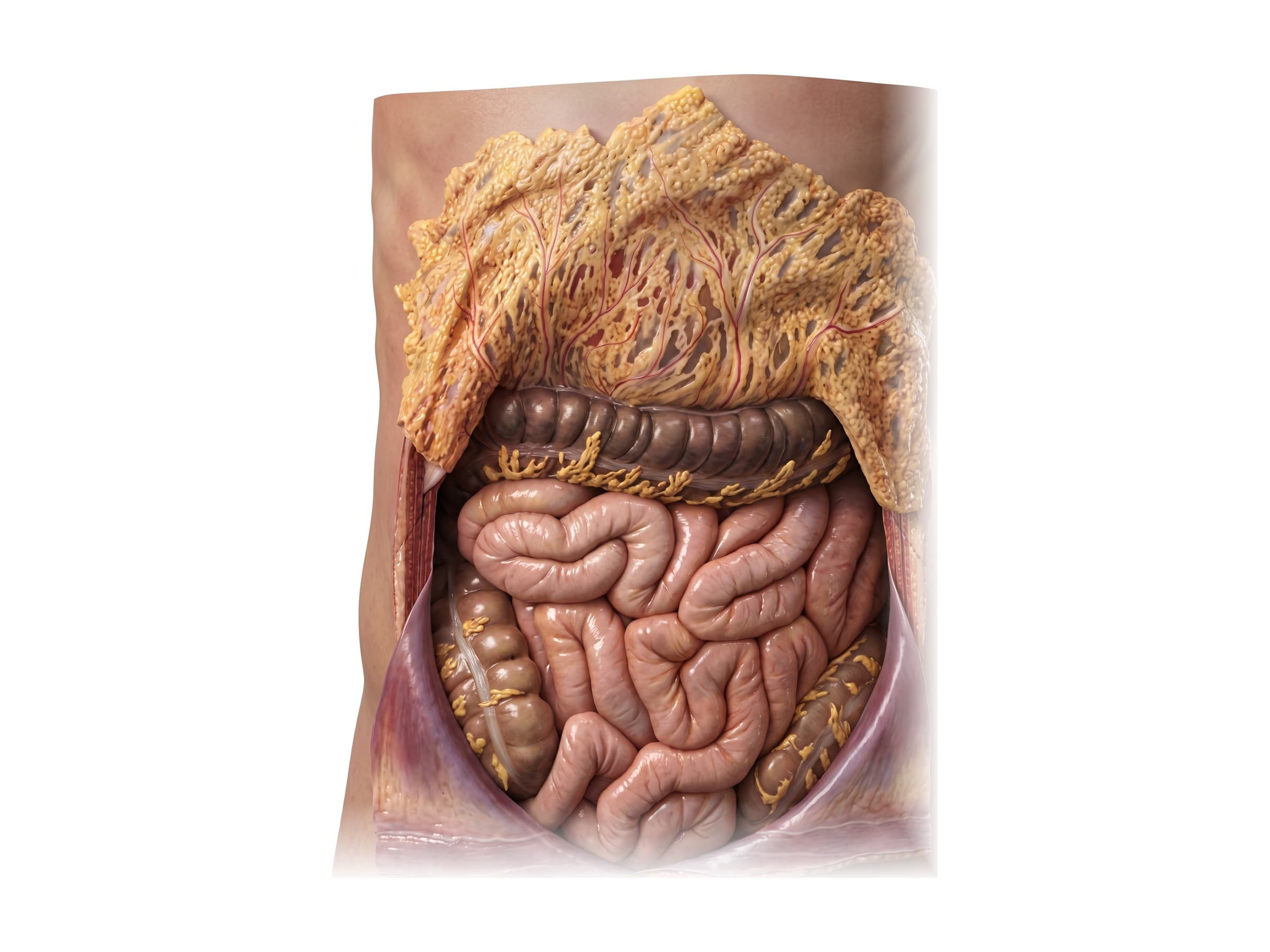
Lifting the omentum up shows the bowel. The transverse colon runs across the top, attached to the back of the omentum. Below it, coiled loops of small intestine fill the middle of the abdomen: the jejunum mostly in the upper left and the ileum mostly in the lower right. The ascending colon runs up the right side and the descending colon down the left, so the large intestine frames the small intestine on three sides.
Abdominal quadrants and regions
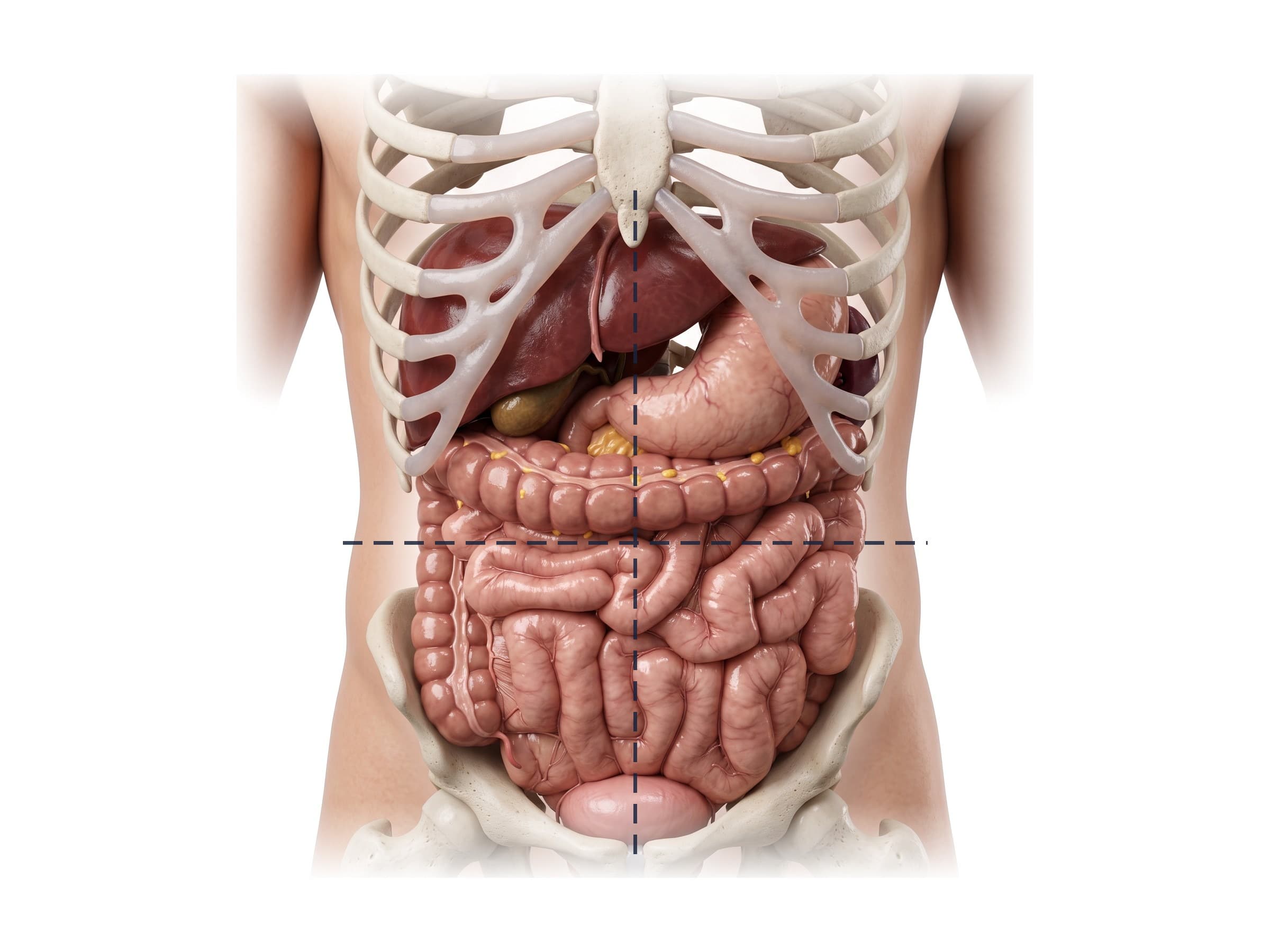
Doctors describe where pain, a mass or a wound is by dividing the abdomen into areas. Right and left always mean the patient's right and left, which is the viewer's left and right in a front view.
- Four quadrants: a vertical line and a horizontal line through the navel give the right upper (RUQ), left upper (LUQ), right lower (RLQ) and left lower (LLQ) quadrants. This is the system used in emergency medicine, imaging reports and the FAST ultrasound exam after trauma.
- Nine regions: two vertical lines through the middle of each collarbone and two horizontal lines (one under the ribs, one between the tops of the hip bones) give the right and left hypochondriac regions under the ribs, the epigastric region in the upper middle, the right and left lumbar (flank) regions, the umbilical region around the navel, the right and left iliac (inguinal) regions, and the hypogastric (suprapubic) region above the pubic bone.
Organs by quadrant:
- Right upper quadrant: most of the liver, gallbladder, duodenum, head of the pancreas, right kidney and adrenal gland, hepatic flexure of the colon.
- Left upper quadrant: stomach, spleen, body and tail of the pancreas, left kidney and adrenal gland, splenic flexure of the colon, left lobe of the liver.
- Right lower quadrant: cecum, appendix, end of the ileum, ascending colon, right ureter.
- Left lower quadrant: descending and sigmoid colon, left ureter.
Below the lower quadrants, the pelvis holds the bladder, the rectum and the reproductive organs; the female pelvis is shown on the female abdominal anatomy page.
The GI tract in order
The gastrointestinal (GI) tract is one long tube. In the abdomen it runs:
- Esophagus (lower end), passing through the diaphragm.
- Stomach, which stores and churns food.
- Duodenum, the first 25 cm of the small intestine, where bile and pancreatic juice enter.
- Jejunum and ileum, the rest of the small intestine, about 6 m (20 feet) in all, where nutrients are absorbed.
- Cecum and appendix, in the right lower quadrant, where the ileum joins the large intestine at the ileocecal valve.
- Ascending, transverse, descending and sigmoid colon, about 1.5 m (5 feet), which absorb water and form stool.
- Rectum and anal canal, in the pelvis.
The liver, gallbladder and pancreas are accessory digestive organs: they are not part of the tube, but they empty bile and enzymes into the duodenum through the bile duct and pancreatic duct.
Stomach diagram
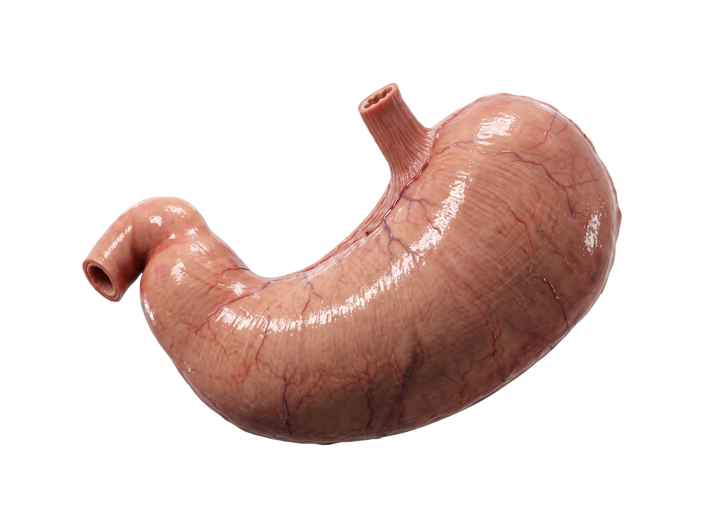
The stomach is a J-shaped bag that sits in the left upper quadrant and the epigastric region, under the left ribs and the left lobe of the liver. Its parts, from top to bottom:
- Cardia: where the esophagus joins the stomach, just below the diaphragm. The lower esophageal sphincter here keeps acid out of the esophagus; when it fails, the result is reflux.
- Fundus: the dome that bulges up to the left of the cardia, under the left half of the diaphragm. It usually holds a bubble of swallowed air, the gastric air bubble seen on a chest X-ray.
- Body: the large middle part, where most of the acid-producing glands are.
- Pyloric antrum: the narrowing lower part that grinds food and pushes it on.
- Pylorus: a thick ring of muscle (the pyloric sphincter) that lets food into the duodenum a little at a time.
The short, concave upper right edge is the lesser curvature, and the long, convex lower left edge is the greater curvature. Inside, the lining is folded into ridges called rugae, which flatten as the stomach fills; an adult stomach holds about 1 to 1.5 liters. Ulcers most often form along the lesser curvature and in the first part of the duodenum, and a perforated ulcer leaks acid and air into the abdomen.
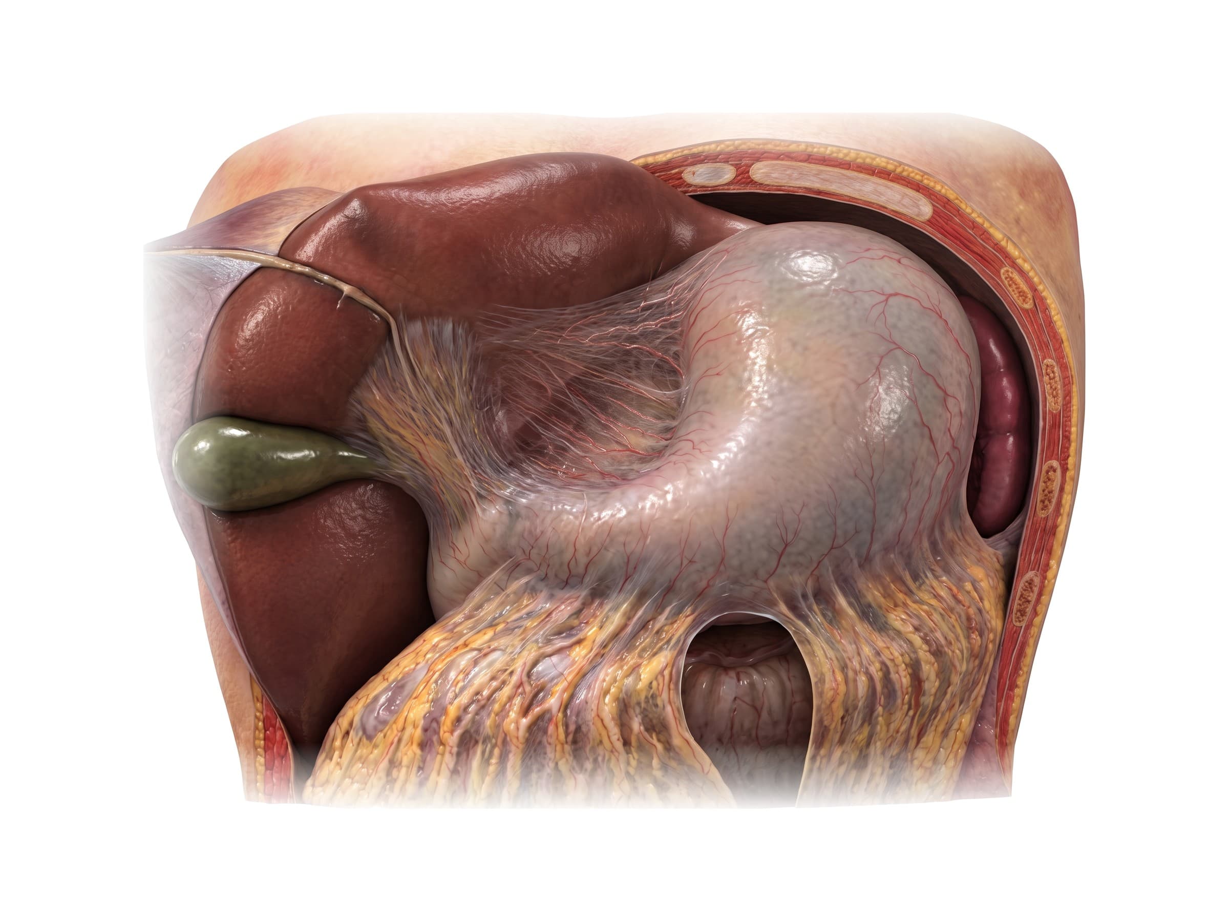
With the liver turned up, the lesser omentum shows as a thin sheet running from the underside of the liver to the lesser curvature of the stomach. Its free right edge, the hepatoduodenal ligament, carries the bile duct, the hepatic artery and the portal vein to the liver. Behind the stomach on the left, the spleen sits against the diaphragm.
Liver and gallbladder
The liver is the largest organ inside the body, about 1.5 kg in an adult. It sits in the right upper quadrant under the diaphragm, protected by the lower ribs, and reaches across the midline into the left upper quadrant. Its main parts are the large right lobe and the smaller left lobe, separated at the front by the falciform ligament; the round ligament (the remnant of the fetal umbilical vein) runs in the lower edge of the falciform ligament. Surgeons and radiologists also divide the liver into eight segments (the Couinaud segments) based on its blood supply, which is how a liver laceration or tumor is located on CT.
The liver receives blood from two sources: the portal vein, which brings blood from the intestines, and the hepatic artery. Blood leaves through the hepatic veins into the inferior vena cava. Because it holds so much blood, a torn liver can bleed heavily.
The gallbladder is a pear-shaped sac, about 7 to 10 cm long, tucked under the right lobe. It stores and concentrates bile made by the liver and squeezes it out after a fatty meal. Gallstones that block its outlet cause biliary colic and cholecystitis, felt as right upper quadrant pain.
Pancreas, duodenum, spleen and bile ducts
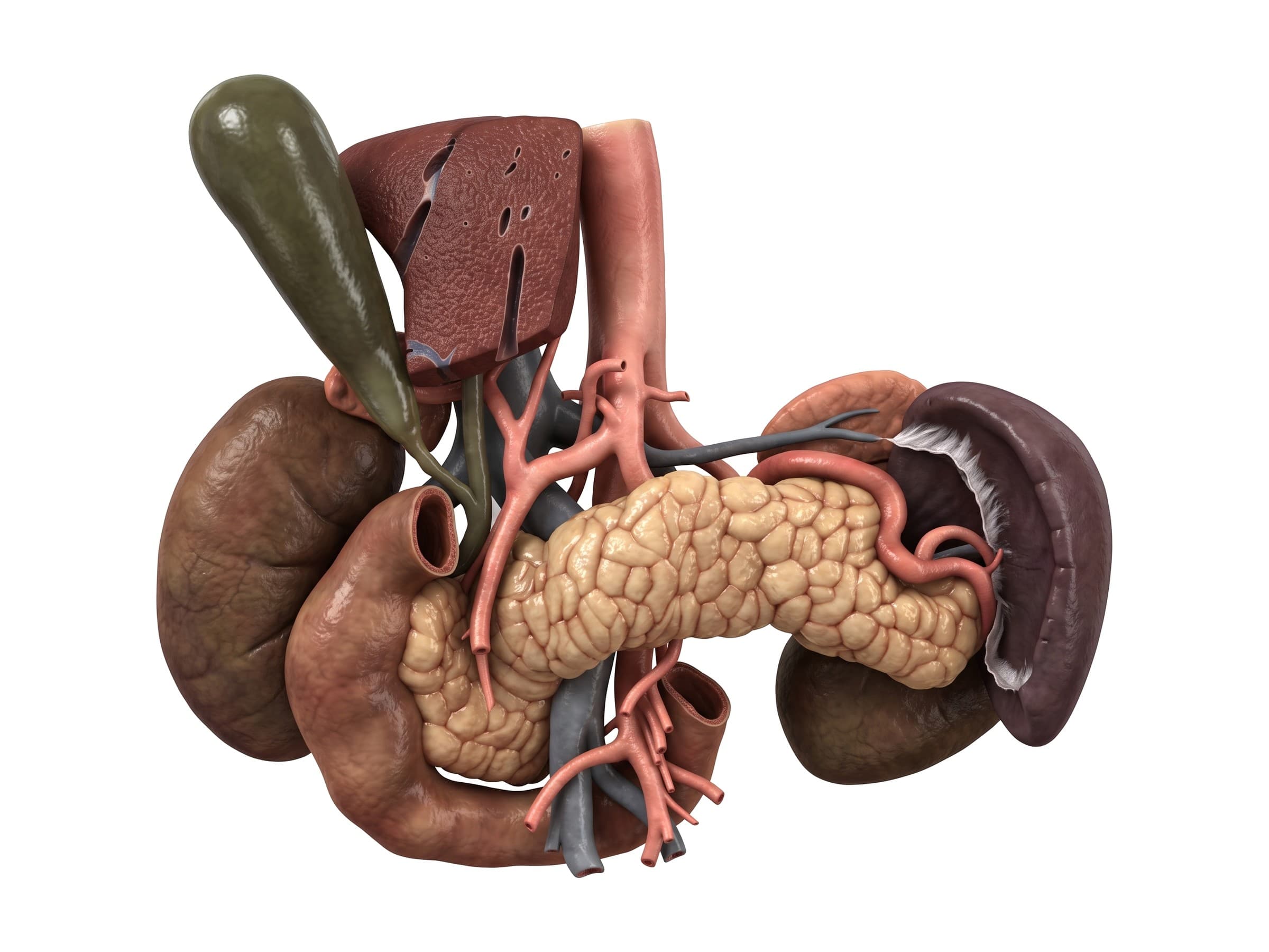
With the stomach removed, the organs of the upper abdomen line up across the back wall:
- Duodenum: the C-shaped first part of the small intestine, about 25 cm long, curving around the head of the pancreas. It has four parts: superior (first), descending (second), horizontal (third) and ascending (fourth). Most of it is retroperitoneal, which is why a duodenal injury from a steering wheel or handlebar can be hard to see at first.
- Pancreas: a soft, lobulated gland about 15 cm long, lying across the spine behind the stomach. Its head sits in the curve of the duodenum, its body crosses in front of the aorta and the superior mesenteric vessels, and its tail reaches the spleen. It makes digestive enzymes (released through the pancreatic duct into the duodenum) and insulin. Because it lies across the spine, a direct blow to the upper abdomen can crush it against the vertebrae.
- Spleen: a fist-sized organ in the left upper quadrant under the 9th to 11th ribs. It filters the blood and helps fight infection, and is the organ most often injured in blunt abdominal trauma.
- Bile ducts: bile drains from the liver through the right and left hepatic ducts into the common hepatic duct. The cystic duct from the gallbladder joins it to form the common bile duct, which runs down behind the first part of the duodenum and through the head of the pancreas to open into the second part of the duodenum, together with the pancreatic duct, at the ampulla of Vater.
The biliary tree matters in surgery: in a cholecystectomy (gallbladder removal), only the cystic duct should be clipped and cut. The anatomy of these ducts varies from person to person, and mistaking the common bile duct for the cystic duct causes a bile duct injury.
Small intestine diagram
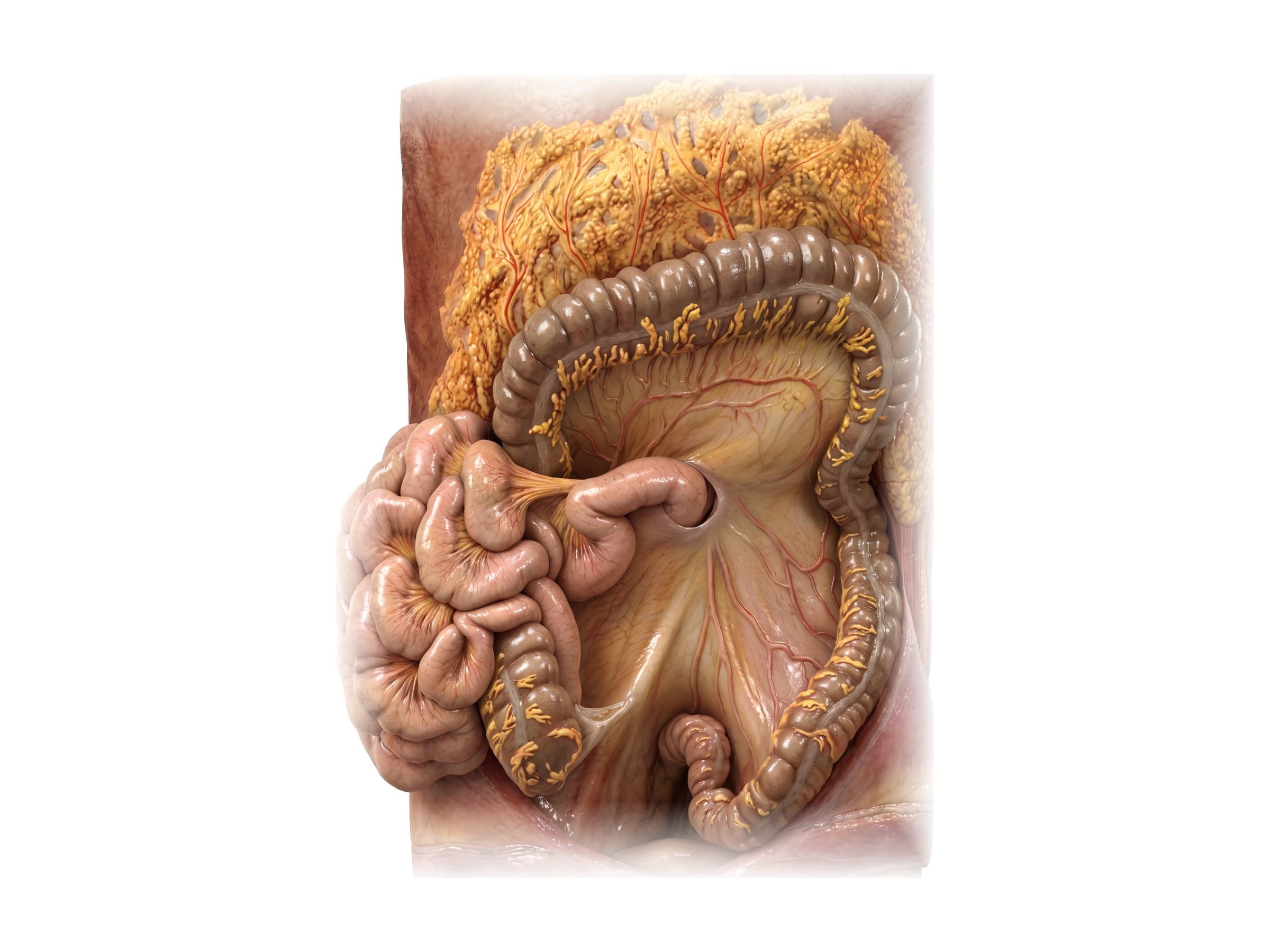
The small intestine, or small bowel, is the longest part of the GI tract. After the duodenum, it surfaces at the duodenojejunal flexure on the left side of the upper abdomen and continues as the jejunum (the first two fifths) and the ileum (the last three fifths), which ends at the cecum in the right lower quadrant. The jejunum is wider, thicker-walled and redder; the ileum is narrower, with more fat in its mesentery.
The jejunum and ileum hang from the back wall on the mesentery, a fan-shaped double fold of peritoneum that carries their blood vessels, lymph nodes and nerves. Its root is only about 15 cm long and runs diagonally from the duodenojejunal flexure, left of the second lumbar vertebra, down to the right sacroiliac joint, but its free edge is as long as the bowel itself. The superior mesenteric artery and vein run in the mesentery; tearing them in a seatbelt injury cuts off the blood supply to a segment of bowel.
Large intestine and colon diagram
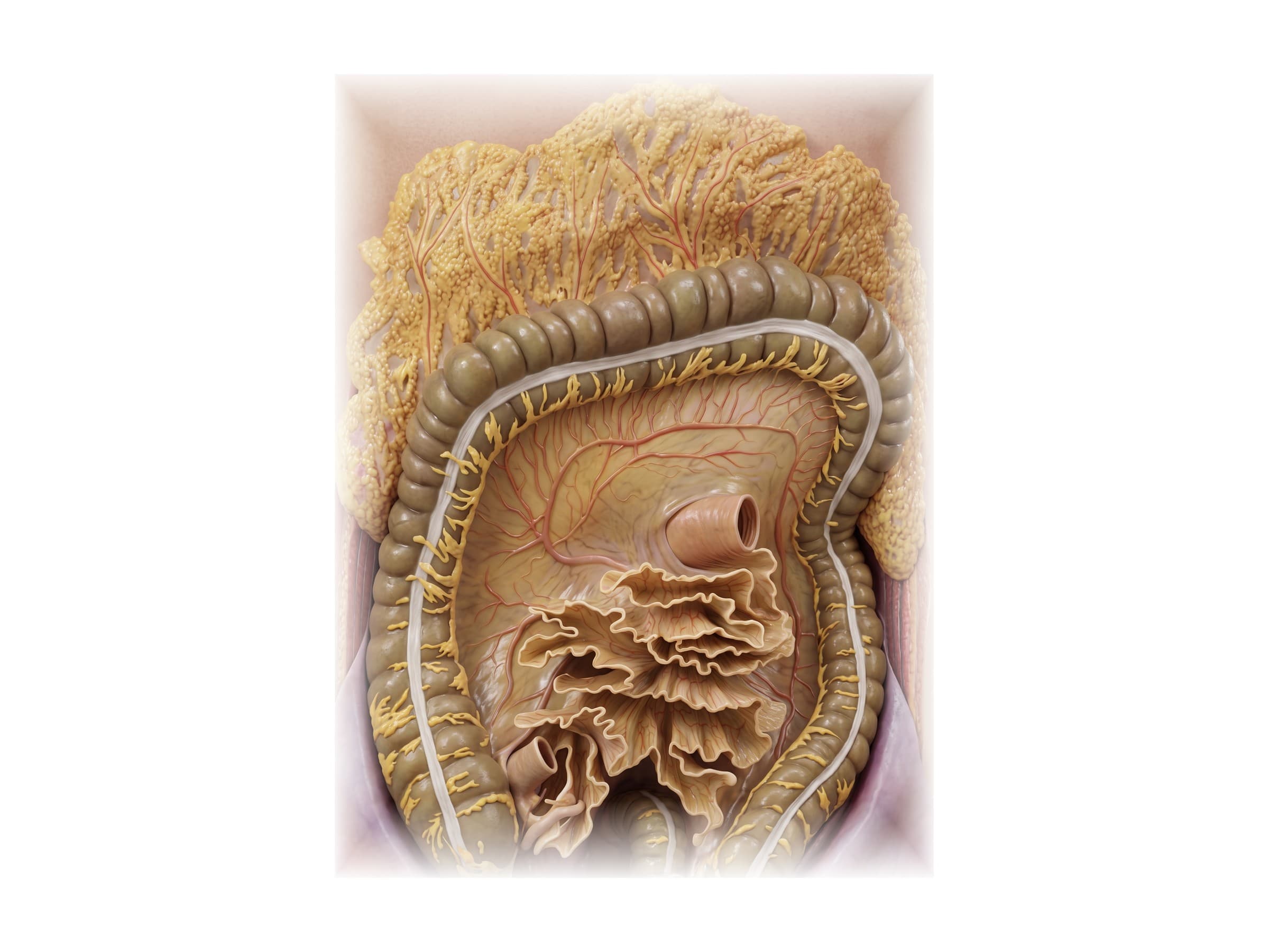
The large intestine, or large bowel, frames the abdomen. It starts at the cecum in the right lower quadrant, climbs the right side as the ascending colon, turns at the hepatic flexure under the liver, crosses the abdomen as the transverse colon (hanging on the transverse mesocolon), turns down at the splenic flexure near the spleen, descends the left side as the descending colon, and curves into the pelvis as the sigmoid colon, which continues as the rectum.
Three features tell the colon apart from the small intestine on a diagram, in surgery or on imaging:
- Taeniae coli: three flat bands of longitudinal muscle running along its length.
- Haustra: the pouches between the bands, which give the colon its segmented look on an X-ray.
- Fatty tags (epiploic appendages) hanging from its surface.
The ascending and descending colon are fixed to the back wall (retroperitoneal), while the transverse and sigmoid colon hang free on a mesentery, so they can twist (volvulus).
Cecum and appendix
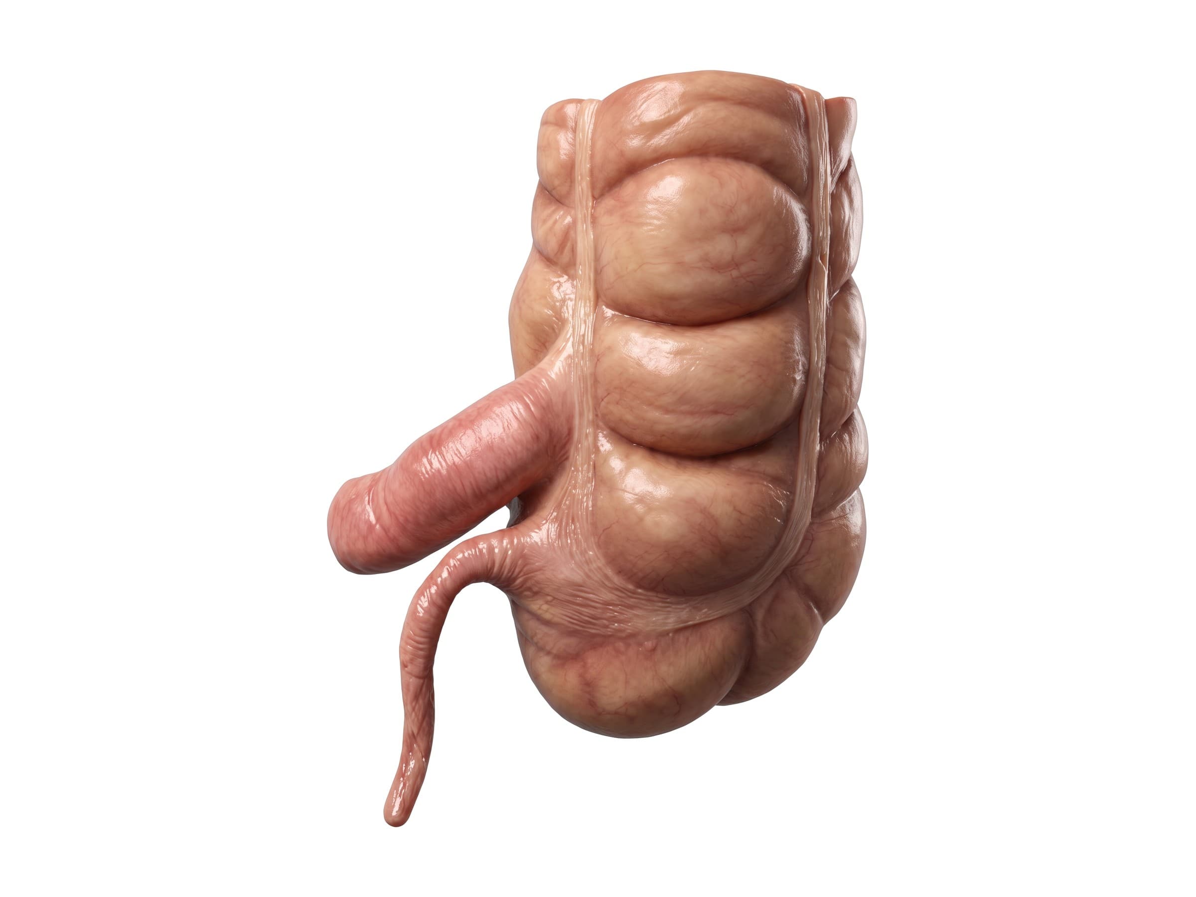
The ileum enters the large intestine at the ileocecal junction, where the ileocecal valve keeps stool from flowing back. Below it, the cecum forms a blind pouch, and the appendix (vermiform appendix), a narrow tube about 8 to 10 cm long, opens into the cecum where the three taeniae coli come together; surgeons follow the bands down to find it. The base of the appendix lies under McBurney's point, one third of the way from the front point of the hip bone to the navel, but its tip can point in many directions: behind the cecum (retrocecal, the most common), down into the pelvis, or next to the ileum. A retrocecal or pelvic appendix gives atypical pain, one reason appendicitis is missed.
Kidneys and the retroperitoneum
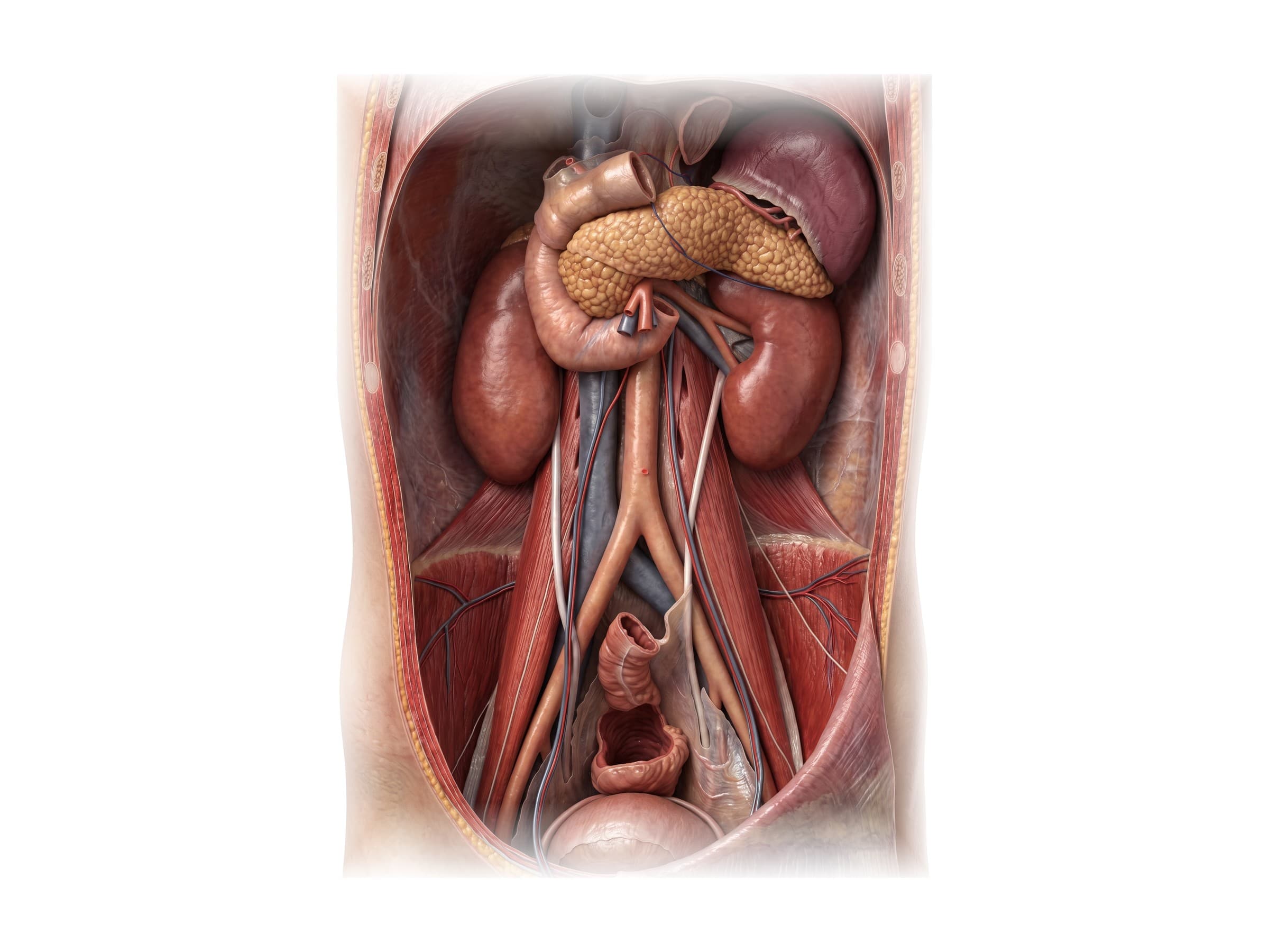
Behind the peritoneum, against the back wall of the abdomen, lies the retroperitoneum. It holds the kidneys and adrenal glands, the ureters, the abdominal aorta and inferior vena cava, most of the duodenum and pancreas, and the ascending and descending colon. These organs lie on the psoas major and quadratus lumborum muscles, on either side of the lumbar spine.
- Kidneys: bean-shaped, about 10 to 12 cm long, from about T12 to L3. The right kidney sits a little lower than the left. The lower ribs cover their upper poles from behind, so a blow to the flank or a fracture of the 11th or 12th rib can bruise or tear a kidney, which shows up as blood in the urine.
- Ureters: each runs down from its kidney on the psoas muscle, crosses the common iliac artery at the pelvic brim and enters the bladder. A kidney stone hurts most where the ureter narrows: where it leaves the kidney, where it crosses the iliac vessels, and where it enters the bladder.
- Aorta and inferior vena cava: the aorta runs down just left of the midline in front of the vertebrae and divides into the two common iliac arteries at about L4; the vena cava runs up on its right. Bleeding into the retroperitoneum from a pelvic or lumbar fracture, or a ruptured aneurysm, can be large and hidden.
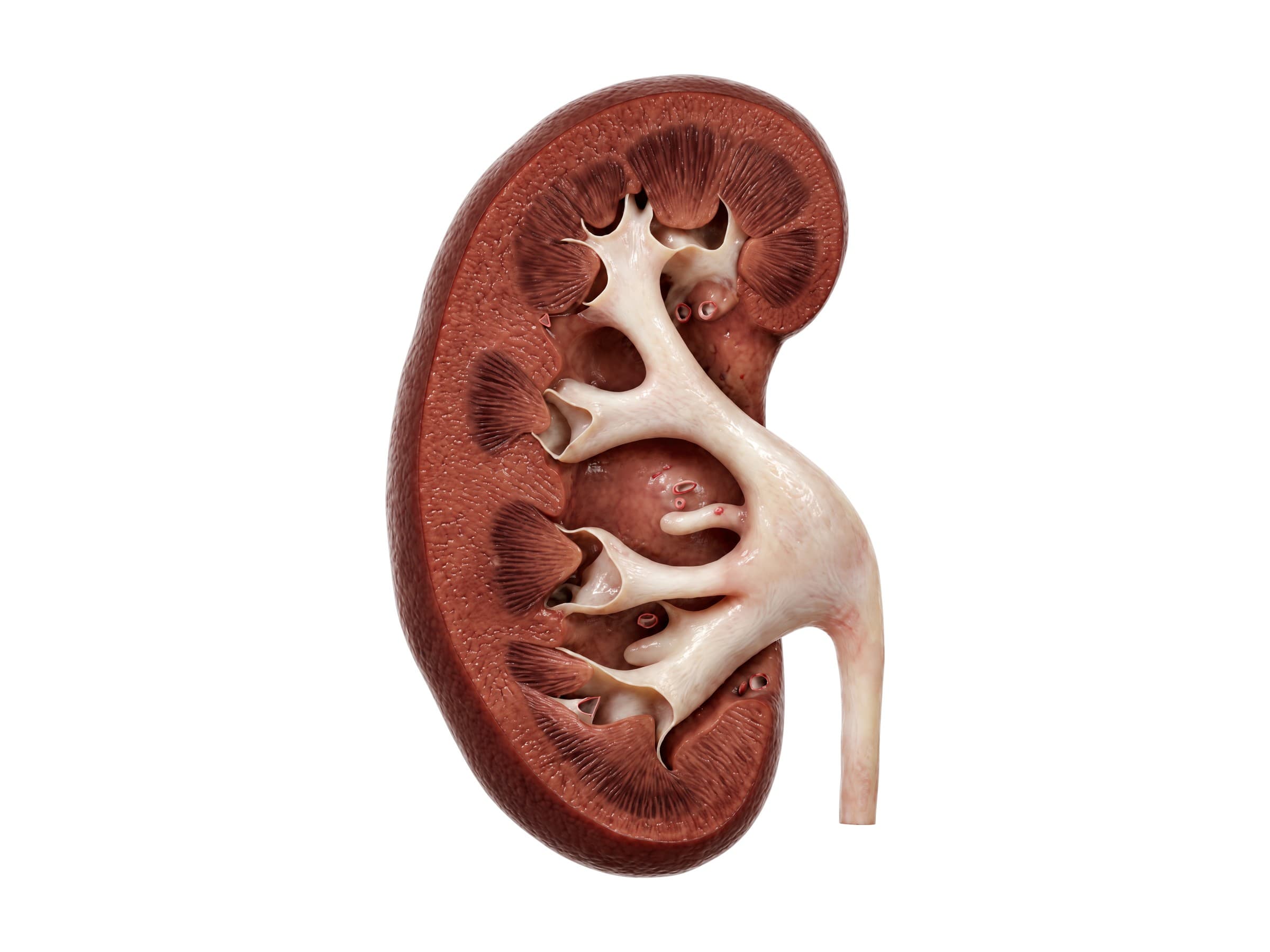
Cut lengthwise, a kidney diagram shows the outer renal cortex, where blood is filtered, and the inner medulla, made of cone-shaped renal pyramids separated by renal columns. Each pyramid ends in a papilla that drips urine into a cup called a minor calyx. Minor calyces join into two or three major calyces, which join into the funnel-shaped renal pelvis in the renal sinus, which narrows into the ureter at the hilum, where the renal artery and vein also enter and leave. Each kidney has about a million filtering units (nephrons).
The peritoneum and omentum
The peritoneum is a thin, slippery membrane. Its parietal layer lines the abdominal wall, and its visceral layer covers the organs, with a thin film of fluid between so the bowel can slide. Organs that hang inside it on a fold of peritoneum (the stomach, most of the small intestine, the transverse and sigmoid colon, the liver and the spleen) are intraperitoneal; organs behind it are retroperitoneal. The greater omentum hangs from the stomach over the bowel, the lesser omentum joins the stomach to the liver, and the mesentery and mesocolons carry blood vessels to the intestines. Blood, bowel contents or infection spreading in this space cause peritonitis, with a rigid, very tender abdomen.
Common injuries and conditions
- Splenic laceration: the most common solid-organ injury in blunt trauma, from car crashes, falls and sports, often with fractures of the lower left ribs. Graded I to V on CT. Bleeding can be delayed: a tear held in by the splenic capsule can rupture hours or days later.
- Liver laceration: the second most common, from blows to the right upper quadrant and fractures of the lower right ribs. Most stop bleeding on their own and are watched in hospital, but a deep laceration can need surgery or embolization.
- Seatbelt injury and bowel perforation: a lap belt pressing into the abdomen in a crash can tear the small intestine, the colon or the mesentery, sometimes with a Chance fracture of the lumbar spine. The seatbelt sign, a bruise across the lower abdomen, is a warning. A perforated bowel leaks its contents into the peritoneum, and the injury can be missed on the first CT scan and show up a day or two later as peritonitis and sepsis.
- Pancreatic and duodenal injury: from a steering wheel, handlebar or a direct blow crushing the upper abdomen against the spine. These retroperitoneal injuries often look minor at first.
- Kidney injury: from a blow to the flank or back; blood in the urine is the main sign. Graded on CT with contrast.
- Missed appendicitis: appendicitis is one of the most common causes of emergency surgery, and a delayed diagnosis lets the appendix rupture, causing peritonitis or an abscess. It is easiest to miss in children, older adults, pregnant women and with a retrocecal or pelvic appendix, and is a common subject of emergency medicine malpractice claims.
- Bile duct injury in cholecystectomy: clipping or cutting the common bile duct instead of the cystic duct during gallbladder removal. If not recognized during the operation, it shows up days later as a bile leak, jaundice or infection, and often needs a major reconstruction.
- Bowel injury in surgery: the bowel can be burned or punctured by a trocar, instrument or energy device during laparoscopy, and a missed injury leads to peritonitis days later.
How abdominal organ injuries are shown in a personal injury or medical malpractice case
Abdominal injuries come up in car crash claims (seatbelt injuries, splenic and liver lacerations, bowel perforation and kidney injuries), falls, assaults and medical malpractice claims involving missed appendicitis, missed bowel injury, and surgical injuries to the bile duct or bowel. A jury cannot see inside the abdomen, so a labeled illustration of the normal anatomy comes first: where the spleen sits under the left ribs, how the lap belt crosses the bowel and mesentery, or how the cystic duct joins the common bile duct. The injury exhibit then builds on the same view: the torn spleen with blood around it, the perforated loop of bowel and the CT findings that were missed, the clip placed across the common bile duct, or the ruptured appendix. Showing the anatomy this way helps a medical expert witness explain why an injury was or was not avoidable, and how it caused the symptoms and surgeries that followed.
Natomy creates custom medical legal illustrations of abdominal injuries for demand packages and trial exhibits. For the rules on using illustrations in court, see demonstrative evidence. Surgeons can use the same figures for patient education materials and in a surgical informed consent discussion before a cholecystectomy, appendectomy or bowel resection. Related atlas pages: female abdominal anatomy and pelvis, lumbar spine anatomy, thoracic spine anatomy, back muscles anatomy, hip anatomy and heart anatomy.
FAQ
What organs are in the abdomen?+
The abdomen holds most of the digestive system: the lower end of the esophagus, the stomach, the small intestine (duodenum, jejunum and ileum), the large intestine (cecum, appendix, colon and upper rectum), the liver, the gallbladder and the pancreas. It also holds the spleen, the two kidneys and adrenal glands, the ureters, and the large blood vessels that run down the back wall, the abdominal aorta and the inferior vena cava. The bladder and the reproductive organs sit below, in the pelvis.
What organs are in each quadrant of the abdomen?+
The right upper quadrant holds most of the liver, the gallbladder, the duodenum, the head of the pancreas, the right kidney and the hepatic flexure of the colon. The left upper quadrant holds the stomach, the spleen, the body and tail of the pancreas, the left kidney and the splenic flexure. The right lower quadrant holds the cecum, the appendix, the end of the ileum and the ascending colon. The left lower quadrant holds the descending and sigmoid colon. Loops of small intestine fill the middle and both lower quadrants. Right and left always mean the patient's side.
What is the difference between the small intestine, the large intestine, the bowel and the colon?+
Bowel and intestine mean the same thing. The small intestine, or small bowel, is about 6 meters (20 feet) long and runs from the stomach to the cecum in three parts: the duodenum, jejunum and ileum. It digests food and absorbs nutrients. The large intestine, or large bowel, is about 1.5 meters (5 feet) long, wider, and runs from the cecum around the edge of the abdomen to the rectum. The colon is the main part of the large intestine (ascending, transverse, descending and sigmoid colon); it absorbs water and forms stool.
What is the GI tract, in order?+
The gastrointestinal (GI) tract is the continuous tube that food passes through: mouth, pharynx, esophagus, stomach, duodenum, jejunum, ileum, cecum (with the appendix), ascending colon, transverse colon, descending colon, sigmoid colon, rectum and anal canal. The liver, gallbladder and pancreas are accessory organs: they are not part of the tube but empty bile and digestive enzymes into the duodenum. The upper GI tract ends at the duodenum, and the lower GI tract begins at the jejunum.
Where are the kidneys located?+
The kidneys lie against the back wall of the abdomen on either side of the spine, roughly from the T12 to the L3 vertebra, behind the peritoneum (retroperitoneal). The right kidney sits a little lower than the left because the liver is above it. The lower ribs cover their upper poles from behind, which is why a blow to the flank or a fracture of the 11th or 12th rib can injure a kidney. Each kidney is about 10 to 12 cm long and drains urine through its ureter to the bladder.
Where is the spleen, and why is it injured so often in car accidents?+
The spleen sits in the left upper quadrant, high up under the 9th to 11th ribs, behind the stomach and against the diaphragm. It is soft, full of blood and held in place by ligaments, so a blow to the left side, a fall or the sudden deceleration of a crash can tear its capsule or split it. The spleen is the abdominal organ most often injured in blunt trauma, and a splenic laceration can bleed heavily into the abdomen. It is graded on CT from I to V and treated with observation, embolization or removal (splenectomy).
What is a seatbelt injury of the abdomen?+
A seatbelt injury happens when a lap belt, or the lap part of a three-point belt, presses into the abdomen in a crash. The bowel and its mesentery are crushed against the spine, which can tear the small intestine, the colon or the mesenteric blood vessels, and the same flexion can fracture the lumbar spine (a Chance fracture). A bruise across the lower abdomen, the seatbelt sign, means a much higher chance of an internal injury. Bowel injuries can look normal on the first CT scan and show up a day or two later as peritonitis.
How does a bile duct injury happen during gallbladder surgery?+
In a laparoscopic cholecystectomy, the surgeon clips and cuts the cystic duct and cystic artery to remove the gallbladder. If the common bile duct or common hepatic duct is mistaken for the cystic duct, which is easier with inflammation, bleeding or unusual anatomy, the main bile duct can be clipped, cut or burned. Bile duct injury happens in roughly 0.3 to 0.5 percent of laparoscopic cholecystectomies, often needs a major reconstruction (hepaticojejunostomy), and is a frequent subject of surgical malpractice claims, which often turn on whether the surgeon confirmed the critical view of safety before cutting.
Need a custom illustration of this joint?
Generate a labeled or exhibit-ready figure for a specific patient, injury, or case in minutes.
Try Natomy →