Sprain vs Strain: Differences, Grades & Healing
A sprain tears a ligament. A strain tears a muscle or tendon. Learn the grades, symptoms, treatment and healing times for each.
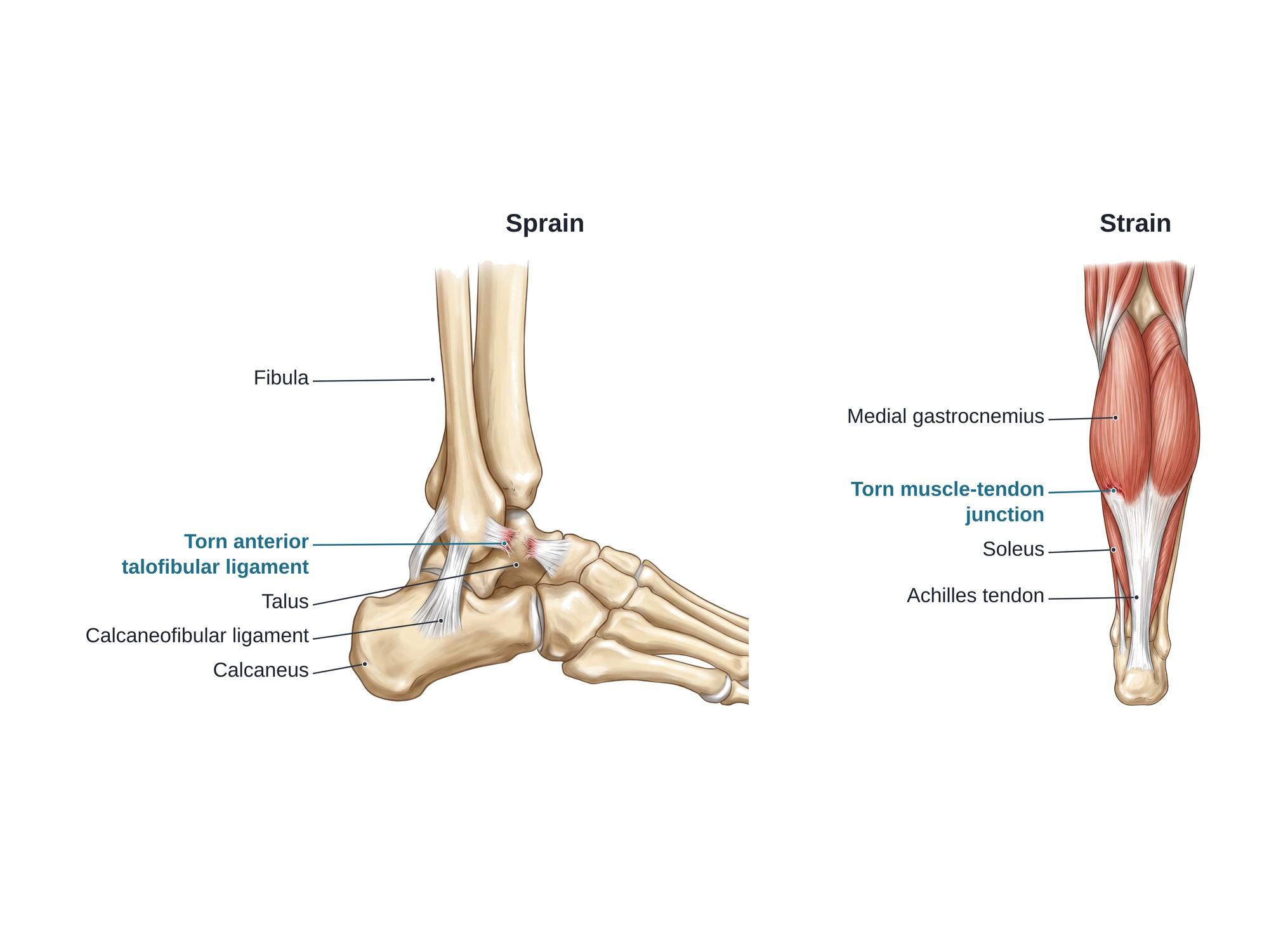
Sprain vs strain comes down to which tissue is torn. A sprain is a stretch or tear of a ligament, the tough band that ties one bone to another at a joint. A strain is a stretch or tear of a muscle or its tendon, the cord that ties muscle to bone. Most strains tear where the muscle fibers meet the tendon, the musculotendinous junction. On the outer ankle, the anterior talofibular ligament (ATFL) runs from the fibula to the talus and the calcaneofibular ligament (CFL) runs down to the heel bone (see ankle anatomy). Both are graded I to III, from a stretch to a complete tear. The most common sprain is a rolled ankle that tears the anterior talofibular ligament. Common strains hit the hamstring and calf muscles. US emergency departments see about 2.15 ankle sprains per 1,000 people each year (Waterman et al., 2010). The figure above shows one of each, side by side.
Sprain vs strain at a glance
| Sprain | Strain | |
|---|---|---|
| Tissue torn | Ligament (bone to bone) | Muscle or tendon (muscle to bone) |
| Usual cause | A joint twisted or bent past its range | A muscle stretched hard while it contracts |
| Common sites | Ankle, knee, wrist, thumb | Hamstring, calf, groin, lower back |
| Where it hurts | Over the joint | In the muscle belly or near the tendon |
| Bruising | Common around the joint | Common, often tracking down the limb |
| Key sign | The joint feels loose or gives way | Pain and weakness when the muscle tightens |
| Main long-term risk | Chronic instability and repeat sprains | Re-tear of the same muscle |
Grades
Both injuries use the same three grades.
- Sprain (ligament)
- Ligament stretched, tiny tears, joint stable
- Strain (muscle or tendon)
- Few fibers torn, strength kept
- Typical healing
- About 1 to 3 weeks
- Sprain (ligament)
- Partial tear, some looseness
- Strain (muscle or tendon)
- Partial tear, clear weakness
- Typical healing
- About 3 to 8 weeks
- Sprain (ligament)
- Complete tear, unstable joint
- Strain (muscle or tendon)
- Complete rupture, often a gap you can feel
- Typical healing
- Several months, sometimes surgery
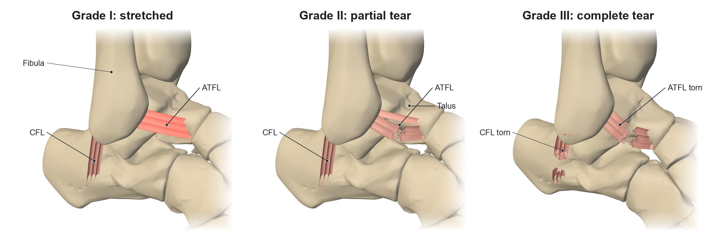
In the ankle, a grade I or II sprain often involves the ATFL alone. A grade III sprain usually tears both the ATFL and the CFL. The figure shows a full-thickness tear of the ATFL with the CFL still intact.
Causes
Sprains happen when a joint is forced past its normal range.
- Rolling the ankle inward (inversion) on uneven ground, a curb or a stair
- Landing on another player's foot in basketball or soccer
- A fall on an outstretched hand, which sprains the wrist
- A twist with the foot planted, which sprains the knee
Lateral sprains make up about 85% of all ankle sprains. The ATFL is the weakest of the outer ligaments and the one most often torn (Ferran and Maffulli, 2006). Ankle sprains peak between ages 15 and 19. About half happen during sports (Waterman et al., 2010).
Strains happen when a muscle is stretched while it is working hard.
- Sprinting, which strains the hamstrings
- Pushing off to jump or lunge, which strains the calf
- Lifting with a twisted or bent back
- Slipping and catching yourself, which can strain the groin or back
In professional soccer, muscle injuries make up 31% of all injuries. The hamstrings account for 37% of them and the calf muscles for 13% (Ekstrand et al., 2011). The calf injury called tennis leg is usually a tear of the medial gastrocnemius, the inner head of the main calf muscle. An ultrasound study found that tear in 66.7% of patients referred with tennis leg (Delgado et al., 2002).
Symptoms
Both cause pain, swelling and bruising. Where and when it hurts is the clue.
Sprain:
- Pain and swelling over the joint, often right after a twist
- A pop at the moment of injury in some cases
- Pain when the joint is moved or loaded
- A loose or unstable feeling with higher grades
Strain:
- Sudden sharp pain in the muscle, sometimes felt as a kick or a snap in the calf
- Pain when the muscle contracts or is stretched
- Muscle spasm and weakness
- A dent or gap in the muscle with a complete tear
Diagnosis
A doctor asks how the injury happened and examines the area. For a sprain, they press on each ligament and test the joint for looseness. The anterior drawer test pulls the foot forward to check the ATFL. For a strain, they look for tenderness along the muscle and test strength against resistance.
X-rays do not show ligaments or muscles. They are used to rule out a fracture. Doctors decide who needs one with the Ottawa ankle rules: bone tenderness at set points, or being unable to bear weight. Ultrasound shows a torn ligament as a thin or broken band with fluid around it. It shows a torn muscle as a gap of fluid at the muscle-tendon junction. MRI shows the full extent of a tear and any bone bruise. See the ankle fracture page for how a break differs.
Treatment
Most sprains and strains heal without surgery.
- Protect and rest the area for the first few days.
- Ice and compression to limit swelling.
- Elevate the limb above the heart.
- Move early. Gentle range of motion starts once pain allows.
- Brace or tape a sprained ankle. A lace-up brace or a walking boot is used for higher grades.
- Rehabilitation. Balance training for sprains. Gradual stretching and loading for strains.
Surgery is uncommon. It is used for a ligament that stays unstable after rehab, often with a Broström repair (tightening the torn ATFL and CFL back to the fibula). It is also used for a tendon pulled off its bone, such as a proximal hamstring avulsion, or a complete tendon rupture. Compare a patellar tendon rupture or an avulsion injury.
Recovery
Recovery depends on the grade. Grade I injuries usually settle in 1 to 3 weeks. Grade II injuries take about 3 to 8 weeks. Grade III injuries take months.
Ligaments heal more slowly than muscle because they have less blood flow. A healed ligament can stay a little longer than before. That is why balance and strength training matter after a sprain. A strain should be loaded gradually. Returning to sprinting too soon is the main reason a hamstring tears again.
Complications
- Chronic ankle instability. About 10% to 30% of people with repeated lateral ankle sprains develop an ankle that keeps giving way (Sarcon et al., 2019).
- Re-injury. In professional soccer, 16% of muscle injuries were re-injuries, and they kept players out longer (Ekstrand et al., 2011).
- Cartilage damage in the ankle joint after a sprain, which can lead to early arthritis
- Scar tissue in a torn muscle, which can make it stiff and prone to tearing again
- Blood clots. A calf injury and time in a boot raise the risk of deep vein thrombosis, which can mimic a calf strain.
- Missed fracture or dislocation. A sprain can hide a fracture, and a joint can partly slip out of place. See subluxation.
How sprains and strains are shown in a personal injury case
Sprains and strains are among the most common injuries in car crashes, slip and falls, and workplace claims. Insurers often call them "just soft tissue." They do not show on X-rays, and that makes them easy to dismiss. A medical illustration shows the jury the exact ligament or muscle that tore, the grade, and why a grade II or III injury can leave a joint unstable or a muscle weak for months. Side-by-side panels, like the figure above, make the difference between a sprain and a strain clear to a lay audience. For how these images are used at trial, see demonstrative evidence and medical legal illustration. A contusion or deeper bruise often occurs with either injury.
References
- Waterman BR, Owens BD, Davey S, Zacchilli MA, Belmont PJ. The epidemiology of ankle sprains in the United States. Journal of Bone and Joint Surgery (American). 2010.
- Ferran NA, Maffulli N. Epidemiology of sprains of the lateral ankle ligament complex. Foot and Ankle Clinics. 2006.
- Ekstrand J, Hägglund M, Waldén M. Epidemiology of muscle injuries in professional football (soccer). American Journal of Sports Medicine. 2011.
- Delgado GJ, Chung CB, Lektrakul N, et al. Tennis leg: clinical US study of 141 patients and anatomic investigation of four cadavers with MR imaging and US. Radiology. 2002.
- Sarcon AK, Heyrani N, Giza E, Kreulen C. Lateral ankle sprain and chronic ankle instability. Foot and Ankle Orthopaedics. 2019.
Related pages
FAQ
How do you know if it's a sprain or a strain?+
Think about what was hurt. A sprain hurts at a joint, often after a twist or a roll, and the joint may feel loose. A strain hurts in the meat of a muscle or where it turns into a tendon, often after a sudden sprint, jump or lift. A strain hurts most when you tighten or stretch that muscle. A doctor confirms it with an exam, and sometimes with ultrasound or MRI.
What heals faster, a sprain or a strain?+
It depends on the grade more than the type. A mild sprain or strain often settles in 1 to 3 weeks. Moderate injuries take several weeks. Muscles have a rich blood supply, so a mild muscle strain may heal a little faster than a ligament. Severe tears of either kind can take months, and some need surgery.
What's worse, an ankle sprain or a strain?+
Neither is always worse. A grade III ankle sprain, a complete ligament tear, can leave a loose, unstable ankle. A grade III strain, a complete muscle or tendon rupture, can cause lasting weakness. Both can be minor at grade I. Ankle sprains are also more likely to happen again.
Do strains heal on their own?+
Most grade I and grade II strains heal without surgery. Rest from painful activity, ice, compression and a guided return to stretching and strengthening help. A complete tear of a tendon, such as a hamstring tendon pulled off the pelvis, may need surgical repair.
Is a pulled muscle a strain?+
Yes. A pulled muscle is the everyday name for a muscle strain. The muscle or its tendon has been stretched too far and some fibers have torn.
Can you walk on a sprained ankle?+
Often you can, with a mild sprain. If you cannot take four steps right after the injury, or there is bone tenderness at the back edge of the ankle bones, get an X-ray to check for a fracture. Walking as pain allows, with a brace, usually speeds recovery after a fracture is ruled out.
Is a sprain worse than a fracture?+
Not always. A fracture is a break in a bone, and a sprain is a ligament tear. A severe sprain can take as long to heal as a simple fracture and can leave the joint unstable. The two often look alike at first, which is why doctors use clear rules to decide who needs an X-ray.
How long does a calf strain take to heal?+
A mild calf strain often heals in 1 to 3 weeks. A partial tear of the medial gastrocnemius, often called tennis leg, usually takes several weeks. A larger tear can take months.
Need this illustration for a patient or a case?
Describe the injury or upload a scan and get a clear, labeled medical illustration for a consult, a chart or a case exhibit.
Try Natomy →