Male Reproductive System Diagram: Organs & Anatomy Labeled
Male reproductive system diagram, labeled: prostate, seminal vesicles, vas deferens, testis, epididymis, spermatic cord, urethra and the male pelvic floor.
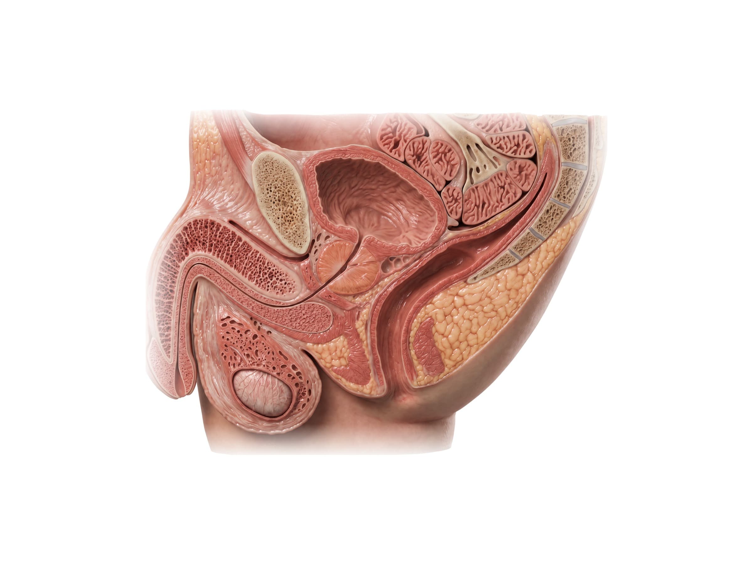
The male reproductive system is the group of organs that make, store and deliver sperm and make the male hormone testosterone. Its parts are the two testes (testicles) in the scrotum, the epididymis on each testis, the vas deferens that carries sperm up through the spermatic cord and inguinal canal into the pelvis, the seminal vesicles and prostate behind and below the bladder, the ejaculatory ducts, the bulbourethral glands, and the urethra running through the penis. Most of these organs sit inside the male pelvis, packed between the pubic bone in front, the bladder above and the rectum behind, which is why prostate, hernia and pelvic surgery can injure the nerves and ducts around them. The figures on this page are labeled medical illustrations of the male reproductive organs and pelvis, for reading imaging and operative reports, explaining a diagnosis or a surgery to a patient, and illustrating an injury or malpractice claim.
Parts of the male reproductive system
Following the path of sperm from where they are made to where they leave the body:
- Testes (testicles): two oval glands in the scrotum that make sperm and testosterone.
- Epididymis: a coiled tube on the top and back of each testis where sperm mature and are stored.
- Vas deferens (ductus deferens): a firm muscular tube that carries sperm from the epididymis up through the spermatic cord, the inguinal canal and the pelvis to the back of the bladder.
- Seminal vesicles: two coiled glands behind the bladder that make most of the fluid in semen.
- Ejaculatory ducts: formed on each side where the vas deferens joins the seminal vesicle duct; they pass through the prostate.
- Prostate: a walnut-sized gland under the bladder that wraps around the urethra and adds a thin, milky fluid to semen.
- Bulbourethral (Cowper's) glands: two pea-sized glands in the pelvic floor that add a clear fluid to the urethra.
- Urethra: the tube that carries both urine and semen out through the penis.
- Penis and scrotum: the external organs. The penis contains three columns of erectile tissue, two corpora cavernosa on top and the corpus spongiosum around the urethra below.
Male pelvis side view (midsagittal section)
The image at the top of this page is a cut straight down the midline of a man's pelvis, with the front of the body on the left. It shows how tightly the reproductive and urinary organs are stacked:
- The urinary bladder sits directly behind the pubic symphysis, with loops of small intestine resting on top of it.
- The prostate lies just under the bladder neck and surrounds the first few centimeters of the urethra.
- The seminal vesicles (cut across in this view) sit behind the bladder and above the prostate, and directly behind them is the rectum, which follows the curve of the sacrum and coccyx.
- The urethra leaves the bottom of the prostate, passes through the pelvic floor and turns forward into the penis inside the corpus spongiosum, while the corpus cavernosum runs above it.
- The testis hangs below in the scrotum, outside the pelvis.
Between the bladder and the rectum the peritoneum dips down to form the rectovesical pouch, the lowest point of the abdominal cavity in a man, where blood or pus collects first. Below it, a thin layer of fascia (Denonvilliers' fascia) separates the prostate and seminal vesicles from the rectum. The same arrangement in a woman is shown on the female abdominal and pelvic anatomy page.
Prostate anatomy and location
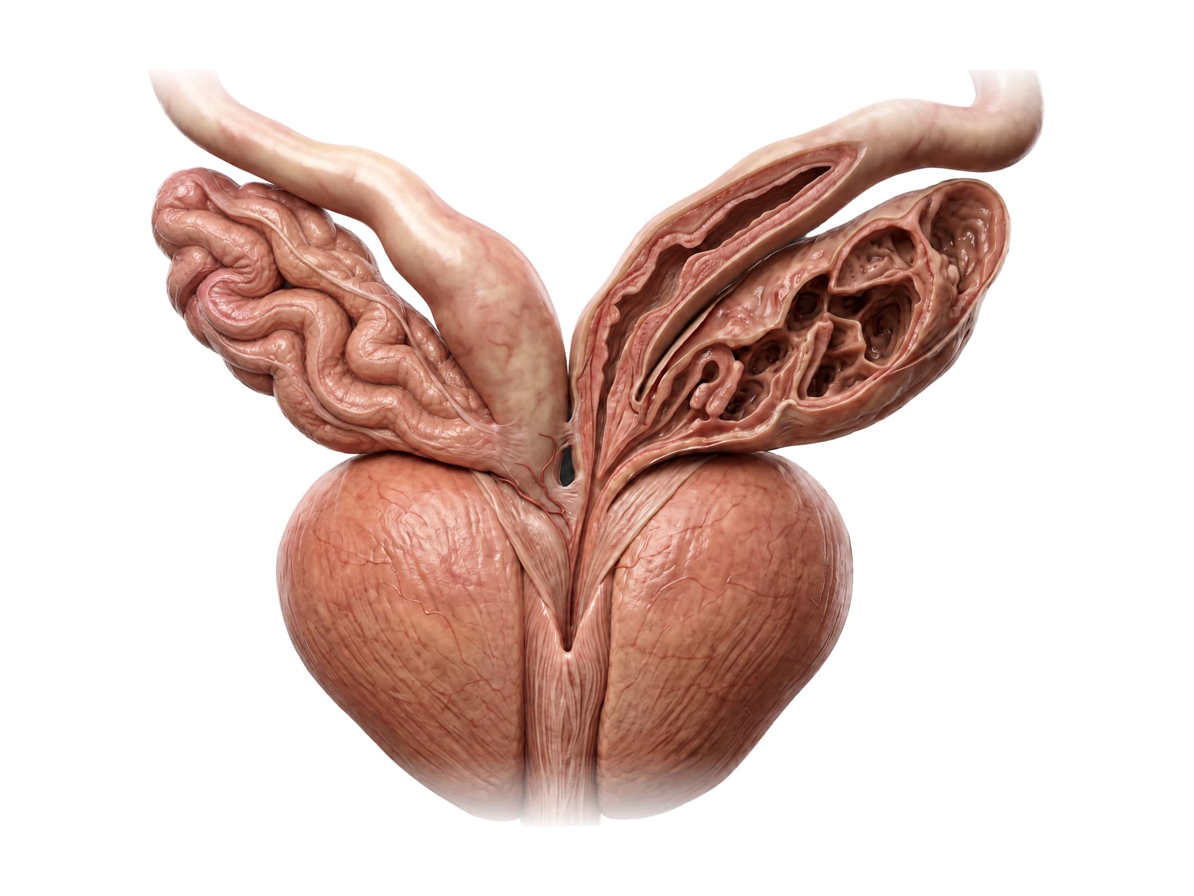
The prostate is a firm, rounded gland about 3 to 4 cm across and about 20 to 25 grams in a young adult, shaped like an upside-down cone. Its wide base is pressed against the bladder neck above and its narrow apex rests on the pelvic floor below. The pubic bone is in front, the rectum is behind, and the levator ani muscles are on each side. Because the back of the prostate lies against the front wall of the rectum, it can be felt on a digital rectal exam and imaged or biopsied through the rectum.
The prostatic urethra runs through the middle of the gland. On its back wall is a ridge, the urethral crest, with a small mound (the verumontanum, or seminal colliculus) where the two ejaculatory ducts open on either side of a tiny blind pocket, the prostatic utricle. Doctors divide the prostate into zones:
- Peripheral zone: the back and sides of the gland, the part felt on rectal exam, where most prostate cancers start.
- Transition zone: around the urethra, the part that enlarges in benign prostatic hyperplasia (BPH) and squeezes the urethra, causing a weak stream.
- Central zone: around the ejaculatory ducts at the base.
- Anterior fibromuscular stroma: a band of muscle and fibrous tissue in front, with no glands.
Seminal vesicles, vas deferens and ejaculatory ducts
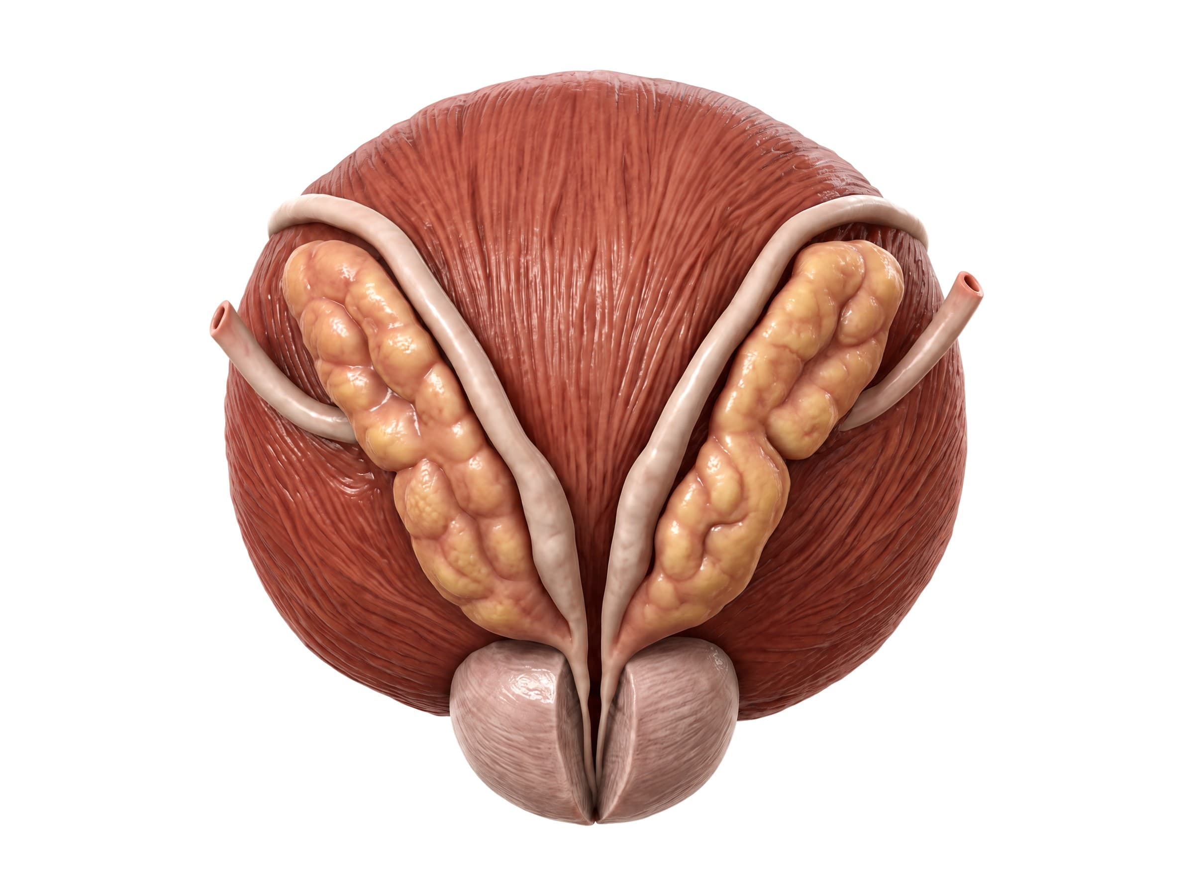
Seen from behind, the back of the bladder carries a V-shaped set of structures. The two seminal vesicles are coiled, lumpy glands about 5 cm long lying on the outside, and the two vasa deferentia run down on the inside of them. Near the bottom each vas deferens widens into a spindle-shaped ampulla, then joins the duct of its seminal vesicle to form an ejaculatory duct, about 2 cm long, which passes through the prostate to the urethra. Higher up, each vas deferens crosses over the ureter just before the ureter enters the bladder.
The seminal vesicles make roughly 60 to 70 percent of the volume of semen, including fructose that fuels sperm, and the prostate adds most of the rest. Their position directly in front of the rectum matters in prostate cancer surgery and radiation, and blockage of the ejaculatory ducts is a treatable cause of male infertility with a low semen volume.
Testicle anatomy: testis and epididymis
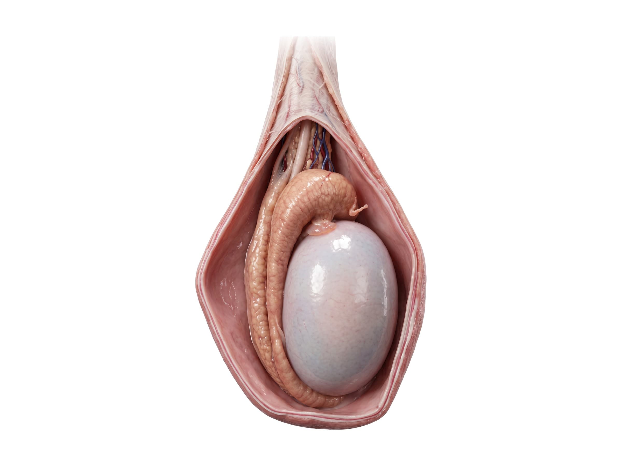
Each testis is an oval gland about 4 to 5 cm long, hanging in the scrotum on its spermatic cord, usually with the left one a little lower. It is wrapped in the tunica vaginalis, a closed two-layered sac of peritoneum left behind when the testis descended from the abdomen before birth. The space inside this sac is where fluid collects in a hydrocele. Normally the tunica vaginalis covers the front and sides of the testis but not its back, which anchors the testis to the scrotal wall. When the tunica wraps all the way around, the testis can swing freely like the clapper of a bell (the bell clapper deformity) and twist on its cord, the cause of most testicular torsion.
The epididymis caps the top and back of the testis. Its head sits on the upper pole, its body runs down the back, and its tail at the lower pole continues as the vas deferens. Small remnants of embryonic ducts, the appendix of the testis and the appendix of the epididymis, hang from the upper pole; they can twist too, causing sudden pain that mimics torsion (a "blue dot sign" under the skin).
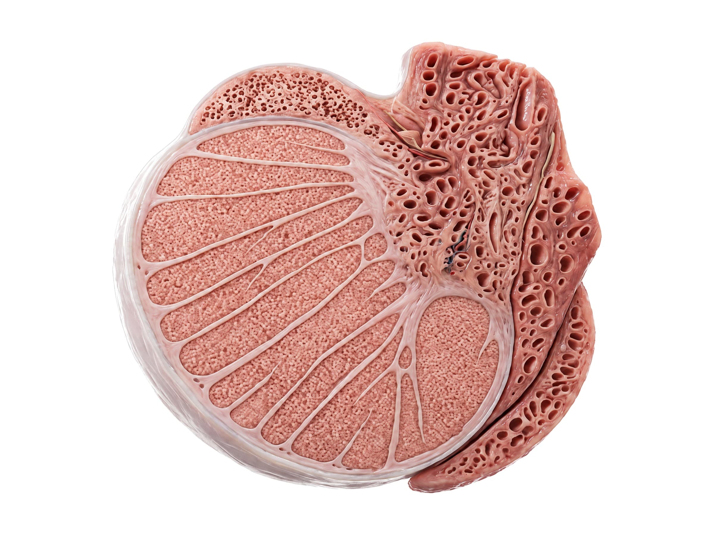
Cut open, the testis is covered by a tough white capsule, the tunica albuginea. Thin walls (septa) run inward from it and divide the testis into about 250 to 300 lobules, each packed with coiled seminiferous tubules where sperm are made over about 2 to 2.5 months. Between the tubules, Leydig cells make testosterone. The tubules drain into a network in the mediastinum testis on the back of the gland and from there into the head of the epididymis. The tunica albuginea is what tears in a testicular rupture after blunt trauma, and its integrity on ultrasound is what decides whether surgery is needed.
Spermatic cord and inguinal canal
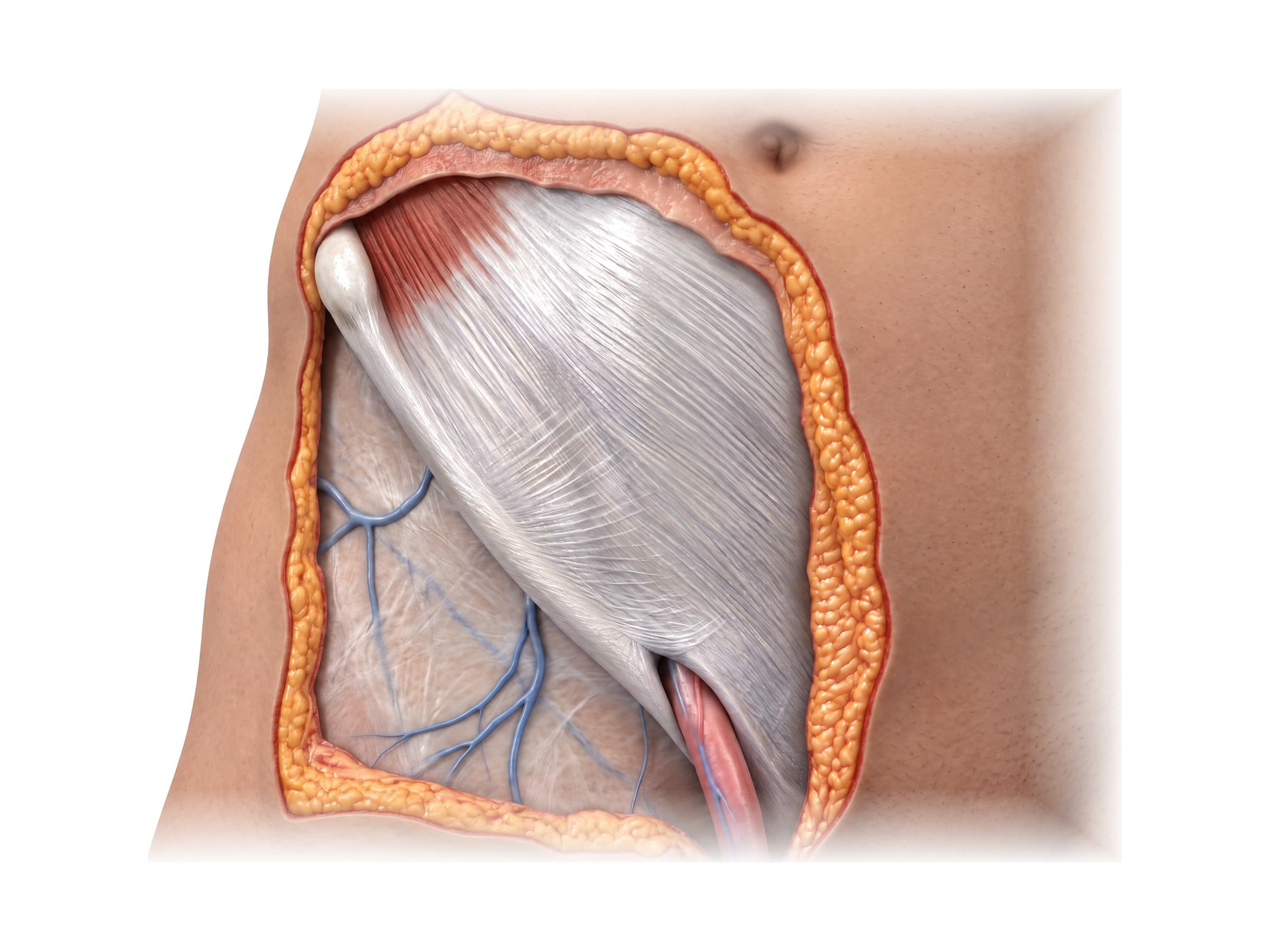
The spermatic cord carries the vas deferens and the testicular vessels between the abdomen and the testis. It starts at the deep inguinal ring, an opening in the transversalis fascia just outside (lateral to) the inferior epigastric artery, runs down and inward through the inguinal canal, a passage about 4 cm long just above the inguinal ligament, and leaves through the superficial inguinal ring to reach the scrotum. Its contents are:
- the vas deferens, which feels like a firm cord when the spermatic cord is rolled between the fingers
- the testicular artery, a long thin branch straight from the aorta, plus the artery to the vas and the cremasteric artery
- the pampiniform plexus, a network of veins that cools the blood going to the testis; when it swells it forms a varicocele, most often on the left because the left testicular vein drains into the left renal vein
- lymphatic vessels, the genital branch of the genitofemoral nerve and sympathetic nerves
The cord is wrapped in three layers taken from the abdominal wall as the testis descended: the external spermatic fascia, the cremaster muscle, which pulls the testis up (the cremasteric reflex), and the internal spermatic fascia. An indirect inguinal hernia follows the same route down the canal inside these coverings, so in a hernia repair the surgeon has to separate the hernia sac from the vas deferens and testicular vessels.
Path of the vas deferens
From the tail of the epididymis the vas deferens runs up the back of the testis, through the spermatic cord and inguinal canal, and enters the abdomen at the deep inguinal ring. There it hooks around the inferior epigastric artery, crosses over the external iliac vessels, and runs down and back along the side wall of the pelvis. It then crosses over the ureter, passes along the back of the bladder inside the seminal vesicle, widens into the ampulla and ends by joining the seminal vesicle duct to form the ejaculatory duct.
Male urethra anatomy
The male urethra is about 18 to 20 cm long, much longer than a woman's, and is described in parts:
- Preprostatic (bladder neck) urethra: a short segment surrounded by the smooth muscle of the internal sphincter.
- Prostatic urethra: about 3 cm through the prostate, where the ejaculatory ducts and prostate glands open.
- Membranous urethra: the shortest and narrowest part, 1 to 2 cm, passing through the pelvic floor inside the external urethral sphincter, the voluntary muscle that keeps a man dry. It is fixed to the pubic bone and is the part torn in pelvic fractures.
- Spongy (penile) urethra: about 15 cm through the corpus spongiosum, starting in the bulb of the penis (the bulbar urethra, the part crushed in straddle injuries) and ending at the external opening (meatus).
Blood supply and nerves of the male pelvis
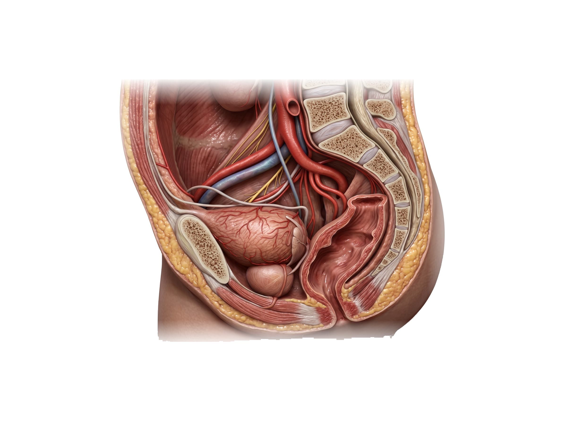
Most male pelvic organs are supplied by branches of the internal iliac artery, which runs down the side wall of the pelvis after the common iliac artery splits. The inferior vesical artery sends branches to the bottom of the bladder, the prostate (prostatic arteries) and the seminal vesicles, and gives the artery to the vas deferens. The internal pudendal artery leaves the pelvis and returns below the pelvic floor to supply the perineum and the erectile tissue of the penis. The testis is different: its testicular artery comes straight off the aorta just below the kidneys and travels the whole length of the spermatic cord, which is why the testis can lose its blood supply when the cord twists or is damaged during groin surgery.
Veins from the prostate form a dense prostatic venous plexus around the gland, including the dorsal venous complex in front, which is a major source of bleeding in prostate surgery. This plexus connects with the veins around the spine, one reason prostate cancer spreads to the lumbar spine and pelvic bones.
The nerves matter most in surgery:
- Cavernous nerves: branches of the pelvic (inferior hypogastric) plexus that carry the parasympathetic signal for erection, from spinal levels S2 to S4. They run in two neurovascular bundles along the back and sides of the prostate, at about the 5 and 7 o'clock positions, then pierce the pelvic floor to reach the corpora cavernosa. Injury to them during radical prostatectomy, rectal cancer surgery or a pelvic fracture causes erectile dysfunction, which is why nerve-sparing prostatectomy dissects the bundles off the prostate.
- Sympathetic nerves (T10 to L2), through the hypogastric nerves, close the bladder neck and drive emission of semen; they can be injured in retroperitoneal lymph node dissection for testicular cancer, causing dry ejaculation.
- Pudendal nerve (S2 to S4): the somatic nerve to the external urethral sphincter, the muscles of the perineum and the skin of the penis and scrotum, and the nerve behind the rhythmic contractions of ejaculation.
The male pelvic floor
The floor of the male pelvis is a sling of muscle, the pelvic diaphragm, made mainly of the levator ani (pubococcygeus, puborectalis and iliococcygeus) with the smaller coccygeus behind it. It runs from the back of the pubic bone and the fascia over the obturator internus on the side wall to the coccyx. In a man only two passages pass through it, the urethra and the anal canal, so the male pelvic floor is stronger and less prone to prolapse than the female one.
The prostate rests directly on the levator ani, with its apex just above the external urethral sphincter in the deep perineal pouch. After a radical prostatectomy the bladder neck sphincter is gone, and continence depends almost entirely on this external sphincter and the surrounding pelvic floor, which is why urinary leakage after prostate surgery is common at first and why pelvic floor exercises are part of recovery. Below the floor, the bulbospongiosus and ischiocavernosus muscles cover the root of the penis, and the perineal body sits between the bulb of the penis and the anus.
Male vs female pelvis
The bones of the pelvis, the two hip bones (ilium, ischium and pubis) and the sacrum, are shown with the hip joint on the hip anatomy page. Compared with the female pelvis described on the female abdominal and pelvic anatomy page, the male pelvis is:
- Narrower and deeper, with taller, more upright iliac wings.
- Heart-shaped at the inlet (android), instead of round or oval.
- Narrow at the pubic arch, with a subpubic angle of about 60 degrees instead of 80 to 90.
- Narrower at the outlet, with the ischial spines and ischial tuberosities closer together.
- Longer and narrower at the sacrum, which is more curved.
- Heavier and rougher, with larger hip sockets (acetabula) and oval rather than triangular obturator foramina.
Inside, the main difference is what sits between the bladder and the rectum: the seminal vesicles, vas deferens and prostate in a man, and the uterus, cervix and vagina in a woman.
Common injuries and conditions
- Testicular torsion: the testis twists on its cord and loses its blood supply, usually in adolescents with a bell clapper deformity. Sudden severe scrotal pain, a high-riding horizontal testis, vomiting and an absent cremasteric reflex are the classic signs. Most testes survive if the cord is untwisted within about 6 hours, and most are lost after 24 hours, so a missed or delayed diagnosis in the emergency department, often labeled epididymitis or a groin strain, is a classic malpractice claim.
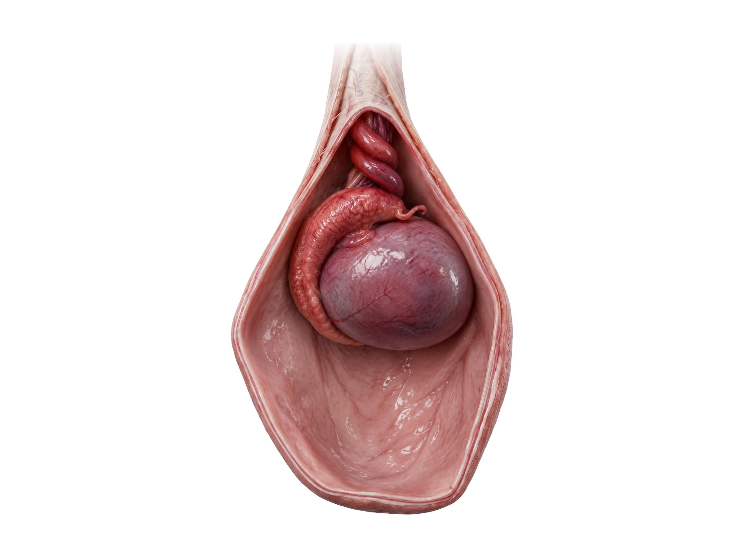
- Hydrocele: clear fluid collects in the tunica vaginalis around the testis, causing a painless, smooth swelling of the scrotum. It is common in newborns, where most go away by age 1 to 2, and in adults it can follow an injury, an infection or surgery such as hernia repair. Ultrasound confirms it and rules out a tumor; a large or bothersome hydrocele is removed surgically (hydrocelectomy).
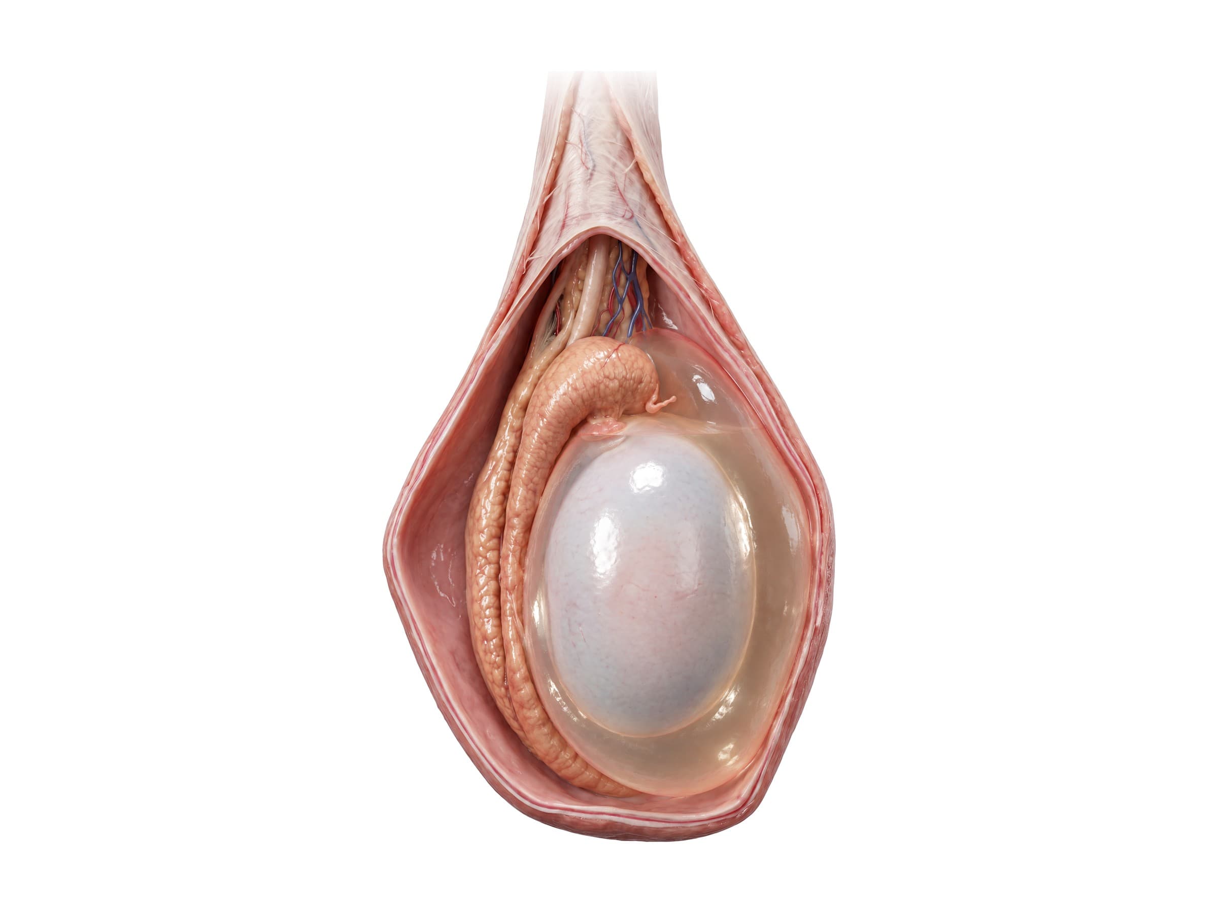
- Testicular rupture and blunt scrotal trauma: a kick, sports injury or crash can tear the tunica albuginea. Ultrasound shows the break, and early surgical repair saves most testes.
- Prostatectomy complications: radical prostatectomy for cancer can injure the cavernous nerves (erectile dysfunction), the external sphincter (urinary incontinence), the rectum behind the prostate (a rectal injury or rectourethral fistula) and, rarely, the ureters at the bladder. Claims usually turn on whether nerve-sparing was appropriate, whether an injury was recognized during surgery, and whether the risks were explained in the consent discussion.
- Vasectomy complications: scrotal bleeding (hematoma), infection, chronic scrotal pain (post-vasectomy pain syndrome), and failure, when the cut ends rejoin or the wrong structure is divided. Pregnancy after vasectomy, especially when the patient was not told to wait for a semen analysis showing no sperm, is a frequent claim.
- Vas deferens injury in inguinal hernia repair: the vas can be cut, crushed, burned or trapped in mesh where it runs with the hernia sac in the inguinal canal. Damage to the testicular artery or pampiniform plexus can cause ischemic orchitis and a shrunken testis, and nerve entrapment causes chronic groin pain. A bilateral injury, or an injury in a man with only one working testis, can cause infertility.
- Urethral injury in pelvic fracture: a crushing pelvic ring fracture can shear the membranous urethra off the prostate where it is fixed to the pubic bone. Blood at the urethral opening, inability to urinate and a high-riding prostate are warning signs, and a retrograde urethrogram should come before a urinary catheter, because forcing a catheter can turn a partial tear into a complete one. Long-term results include urethral stricture, incontinence and erectile dysfunction.
- Straddle injury: a fall astride a bar, beam or bicycle crushes the bulbar urethra against the underside of the pubic bone, causing bruising, bleeding and later a stricture.
- Fournier's gangrene: a fast-spreading necrotizing infection of the scrotum, perineum and penis, more common in men with diabetes, obesity or recent perineal surgery. Early signs (pain out of proportion to what is seen, swelling, fever) can look like cellulitis or an abscess, and every hour of delay before surgical debridement raises the risk of death, so a missed diagnosis is a recurring claim.
- Benign prostatic hyperplasia and prostate cancer: enlargement of the transition zone squeezes the urethra and causes urinary symptoms in older men, while most cancers start in the peripheral zone and are found by PSA testing, rectal exam, MRI and biopsy.
How male reproductive anatomy is shown in a personal injury or medical malpractice case
Male reproductive anatomy comes up in medical malpractice claims involving urology and general surgery (a missed testicular torsion, a prostatectomy with nerve or sphincter injury, a failed or complicated vasectomy, a vas deferens or testicular artery injury during hernia repair, a delayed diagnosis of Fournier's gangrene) and in personal injury claims involving pelvic fractures, straddle falls and blunt scrotal trauma. Few jurors know where the prostate is, that the testis gets its blood from a single long artery in the spermatic cord, or that the nerves for erection run in thin bundles along the side of the prostate. A labeled illustration of the normal anatomy comes first: the midline section of the pelvis, the spermatic cord passing through the inguinal canal, or the neurovascular bundles beside the prostate. The injury exhibit then builds on the same view: the twisted cord and dark, swollen testis, the cut vas deferens next to the hernia mesh, the clipped nerve bundle, or the urethra torn off the prostate by a displaced pelvic fracture. Showing the anatomy this way helps the medical expert witness explain why an injury was or was not avoidable, and why it caused the impotence, incontinence, infertility or loss of a testis that followed.
Natomy creates custom medical legal illustrations of pelvic, urologic and groin injuries for demand packages and trial exhibits. For the rules on using illustrations in court, see demonstrative evidence. Urologists and general surgeons can use the same figures in a surgical informed consent discussion before a prostatectomy, vasectomy or hernia repair. Related atlas pages: female abdominal and pelvic anatomy, abdominal organs, hip anatomy, lumbar spine anatomy and leg nerves and dermatomes.
FAQ
What are the parts of the male reproductive system?+
The male reproductive system has internal and external parts. The two testes (testicles) make sperm and testosterone and sit in the scrotum. Each testis drains into an epididymis, where sperm mature, and then into a vas deferens, which travels up through the spermatic cord and inguinal canal into the pelvis. Behind the bladder each vas deferens joins a seminal vesicle to form an ejaculatory duct, which passes through the prostate and opens into the urethra. The urethra then carries semen (and urine) through the penis. The bulbourethral glands add a small amount of fluid near the start of the penile urethra.
Where is the prostate located?+
The prostate sits deep in the pelvis, directly below the bladder and in front of the rectum, behind the lower edge of the pubic bone. It wraps around the first part of the urethra as it leaves the bladder. Because its back surface lies against the front wall of the rectum, a doctor can feel the prostate with a gloved finger during a digital rectal exam. Its lower tip (apex) rests on the pelvic floor, just above the external urinary sphincter.
What does the vas deferens do?+
The vas deferens (ductus deferens) is a muscular tube about 30 to 45 cm long that carries sperm from the tail of the epididymis to the ejaculatory duct. It runs up inside the spermatic cord, through the inguinal canal, along the side wall of the pelvis and over the ureter to the back of the bladder, where it widens into the ampulla. During ejaculation its thick muscular wall contracts to push sperm forward. Cutting or blocking both vasa deferentia, as in a vasectomy, stops sperm from reaching the semen.
What is the epididymis?+
The epididymis is a tightly coiled tube, about 6 meters long if straightened, that lies along the top and back of each testis. It has a head at the top, which receives sperm from the testis, a body along the back, and a tail at the bottom, which continues as the vas deferens. Sperm spend roughly two weeks passing through it, gaining the ability to swim and fertilize an egg. Inflammation of the epididymis (epididymitis) is a common cause of scrotal pain and has to be told apart from testicular torsion.
What do the seminal vesicles do?+
The seminal vesicles are two coiled glands, each about 5 cm long, that lie behind the bladder and above the prostate, just outside each vas deferens. They make most of the fluid in semen, roughly 60 to 70 percent of its volume, including fructose that feeds the sperm. Each seminal vesicle duct joins the ampulla of the vas deferens to form an ejaculatory duct, which runs through the prostate to open into the urethra.
What is in the spermatic cord?+
The spermatic cord runs from the deep inguinal ring in the lower abdominal wall down to the back of the testis. It contains the vas deferens, the testicular artery (straight from the aorta), the artery to the vas deferens, the cremasteric artery, the pampiniform plexus of veins, lymphatic vessels, the genital branch of the genitofemoral nerve and sympathetic nerves. It is wrapped in three layers taken from the abdominal wall: the external spermatic fascia, the cremaster muscle and the internal spermatic fascia.
Why is testicular torsion an emergency?+
In testicular torsion the testis twists on its spermatic cord and cuts off its own blood supply. It is most common in adolescents, often in boys whose testis hangs freely inside the tunica vaginalis (a bell clapper deformity). Most testes are saved if the cord is untwisted within about 6 hours, while after 24 hours most are lost. Sudden severe scrotal pain, a high-riding testis, nausea and an absent cremasteric reflex should lead to an urgent urology consult and Doppler ultrasound, and surgery should not be delayed for imaging when the clinical picture is clear.
How can prostate surgery cause erectile dysfunction?+
The cavernous nerves, which control erection, run in two thin neurovascular bundles along the back and sides of the prostate, at about the 5 and 7 o'clock positions, before passing through the pelvic floor to the erectile tissue of the penis. During a radical prostatectomy the surgeon removes the prostate from between these bundles. If the nerves are cut, burned, stretched or tied, erections can be weakened or lost. Nerve-sparing techniques peel the bundles off the prostate to protect them, but cancer location sometimes requires removing one or both.
Need a custom illustration of this joint?
Generate a labeled or exhibit-ready figure for a specific patient, injury, or case in minutes.
Try Natomy →