Comminuted Fracture: Definition, X-ray & Treatment
A comminuted fracture is a bone broken into three or more pieces. See a comminuted hip fracture on X-ray beside a labeled medical illustration.
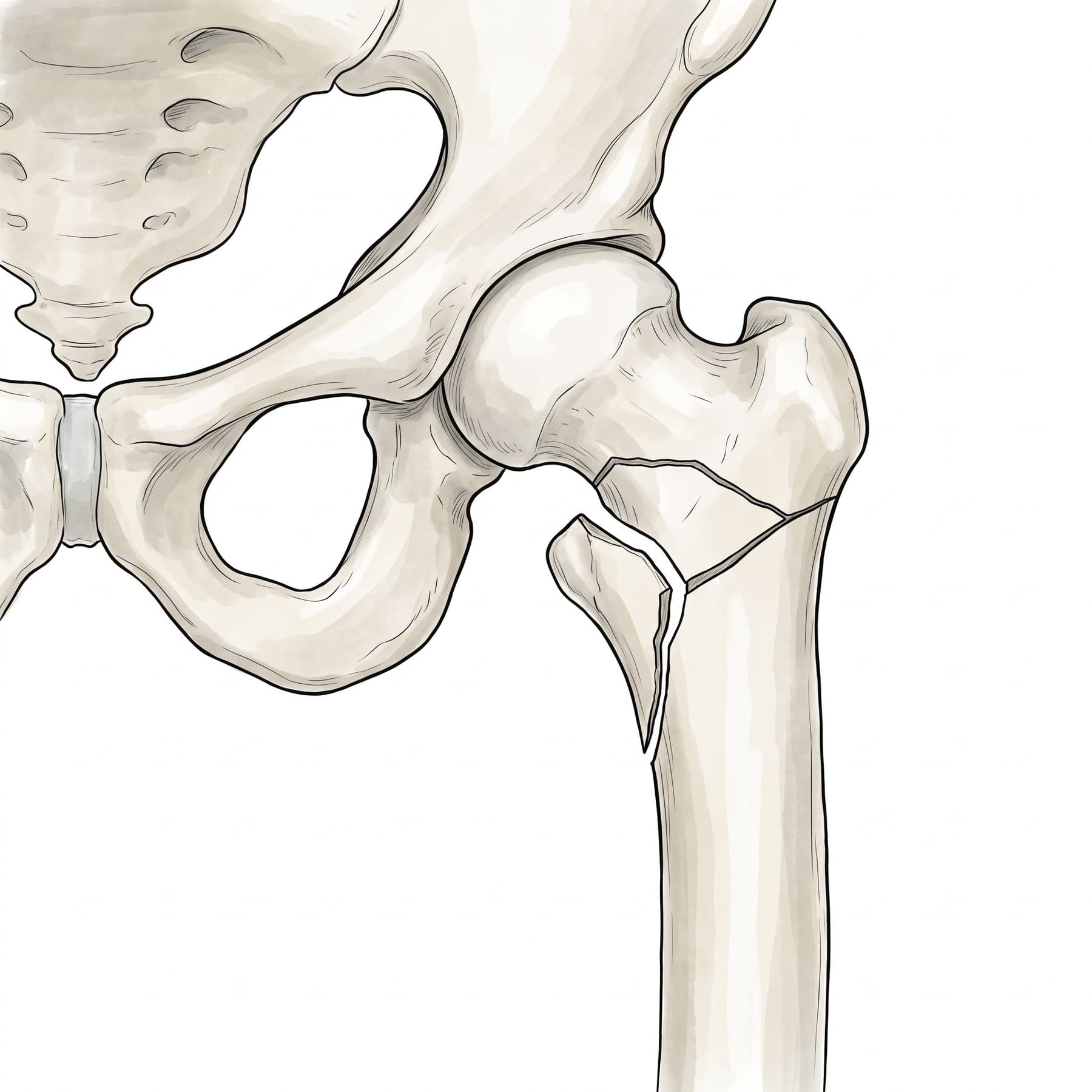
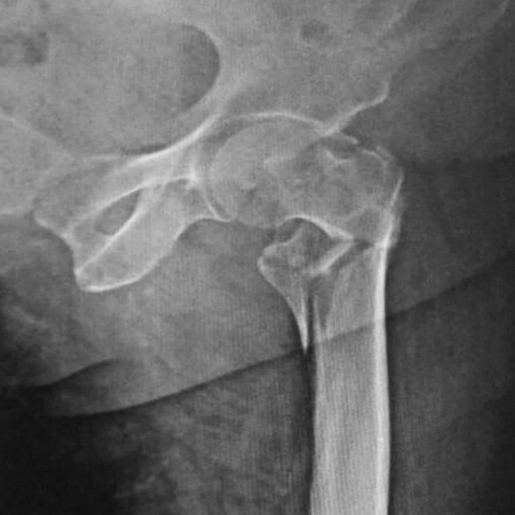
A comminuted fracture is a break in which a bone is broken into three or more pieces, instead of the two pieces of a simple fracture. The word comes from the Latin for "broken into small pieces." Comminuted fractures usually take more force, are less stable, and more often need surgery. The figure above shows a real X-ray of a comminuted hip fracture with a medical illustration drawn from it: drag the slider to compare them.
What makes a fracture comminuted?
Fractures are described by how many pieces the bone breaks into:
- Simple fracture: one fracture line and two pieces.
- Wedge (butterfly) fracture: two main pieces plus a wedge-shaped third piece, but the main pieces still touch.
- Comminuted (multifragmentary) fracture: three or more pieces, often with small fragments, so the main pieces no longer touch.
A comminuted fracture can also be displaced (the pieces have moved out of line) or non-displaced, and closed or open (the skin is broken). Each of these changes the treatment. The AO/OTA classification that surgeons use labels simple, wedge and multifragmentary fractures as types A, B and C for most long bones.
Comminuted fracture on X-ray
The X-ray above is a front (AP) view of a left hip. The upper end of the femur is broken between the greater trochanter (the bony point on the outside of the hip) and the lesser trochanter (a smaller knob on the inside) into four pieces: the femoral head and neck still attached to the greater trochanter, a wedge-shaped fragment between the fracture lines, the lesser trochanter broken off on its own, and the shaft. This is a comminuted intertrochanteric fracture, one of the two main types of hip fracture in older adults.
On any X-ray, comminution shows as several fracture lines meeting each other and loose pieces of bone between the main fragments. CT is often added when the fracture enters a joint, because small fragments of the joint surface are hard to count on plain films.
Common comminuted fractures
Any bone can break into several pieces, but some patterns are especially common:
- Hip (intertrochanteric) fractures, like the one above, in older adults after a fall.
- Distal radius fractures at the wrist, including many Colles fractures.
- Tibial plateau and pilon fractures, where the joint surface of the tibia breaks into several pieces.
- Calcaneus (heel bone) fractures from falls from a height.
- Patella fractures from a direct blow to the kneecap.
- Femoral and tibial shaft fractures from car, motorcycle and pedestrian crashes.
How a comminuted fracture happens
Comminution reflects how much energy the bone absorbed. In younger adults it usually takes a high energy injury: a car or motorcycle crash, a fall from a height, a crush injury or a gunshot wound. In older adults with osteoporosis, the bone is weaker and can break into several pieces from a fall from standing, which is why comminuted hip, wrist and shoulder fractures are common in older patients.
Symptoms
- Severe pain at the injury site
- Swelling and bruising, often extensive
- Deformity, shortening or abnormal rotation of the limb
- Inability to move the limb or bear weight
- An open wound over the fracture in an open (compound) fracture
Treatment
Treatment depends on the bone, the amount of displacement, whether the joint surface is involved, and the patient's health.
- Cast, splint or brace: for stable comminuted fractures with little displacement, especially in the upper limb.
- Internal fixation: plates and screws, or a rod (intramedullary nail) through the center of the bone, to hold the pieces in alignment. Intertrochanteric hip fractures like the one above are usually fixed with a nail or a sliding hip screw.
- Bridge plating: in very comminuted fractures, a plate spans the broken zone without fixing every small piece, restoring length and alignment while the fragments heal around it.
- External fixation: pins and a frame outside the limb, often a first step when the soft tissue is badly injured.
- Joint replacement: when the joint surface is shattered beyond repair, for example in some hip and shoulder fractures in older patients.
Recovery time
Comminuted fractures usually heal more slowly than simple fractures of the same bone. Small bones may heal in 6 to 8 weeks; comminuted fractures of the femur or tibia often take 3 to 6 months. After hip fracture surgery, most patients start walking with a walker within a day or two, but regaining independence can take months. Physical therapy is a large part of recovery.
Complications
- Delayed union or nonunion: the bone heals slowly or not at all
- Malunion: the bone heals shortened, angled or rotated
- Infection, especially after open fractures or extensive surgery
- Post-traumatic arthritis when the fracture enters a joint
- Compartment syndrome after high energy fractures of the leg or forearm
- Blood clots and, after hip fractures in older adults, pneumonia and loss of independence
How a comminuted fracture is shown in a personal injury case
"Comminuted" is a word jurors have never heard, and it is one of the most important in a fracture case: it explains why the injury needed surgery, why recovery took months, and why arthritis or hardware problems may follow. An illustration drawn from the plaintiff's own X-ray, like the one above, shows each fragment clearly, where a black and white film shows a blur of overlapping lines. Typical exhibits pair the comminuted fracture with the plates, screws or nail placed at surgery. For how these images are used at trial, see demonstrative evidence, trial exhibits and medical legal illustration.
Related pages
FAQ
What is a comminuted fracture?+
A comminuted fracture is a break in which the bone is broken into three or more pieces, instead of the two pieces of a simple fracture. It usually takes more force to cause, and it is harder to put back together and hold in place while it heals.
Is a comminuted fracture serious?+
It is generally more serious than a simple fracture. With more pieces, the bone is less stable, more likely to need surgery, and slower to heal, and the surrounding soft tissue is often more damaged. How serious it is depends on the bone, whether the fracture enters a joint, and whether the skin is broken.
Does a comminuted fracture always need surgery?+
Not always. Some comminuted fractures with little displacement, especially in bones like the ribs, fingers or the shaft of the humerus, heal in a cast, brace or sling. Most comminuted fractures of the hip, the joint surfaces and the long bones of the leg are fixed with surgery so the patient can move and bear weight sooner.
How long does a comminuted fracture take to heal?+
Usually longer than a simple fracture of the same bone. Small bones may heal in 6 to 8 weeks, while a comminuted fracture of the femur or tibia often takes 3 to 6 months, and sometimes longer. Recovery of strength and motion can take a year.
What is the difference between a comminuted and a displaced fracture?+
Comminuted describes how many pieces the bone breaks into (three or more). Displaced describes whether the pieces have moved out of line. A fracture can be comminuted and displaced, comminuted and non-displaced, or simple and displaced.
What is the difference between a comminuted and a segmental fracture?+
A segmental fracture is a specific pattern in which the bone breaks in two separate places, leaving a free middle segment. A comminuted fracture breaks into three or more pieces at one area. Segmental fractures are sometimes counted as a type of multifragmentary fracture.
What causes a comminuted fracture?+
High energy injuries such as car and motorcycle crashes, falls from a height and crush injuries are the classic causes. In older adults with osteoporosis, a simple fall from standing can be enough to break the hip, wrist or shoulder into several pieces.
Need an illustration of a patient's own X-ray?
Upload an X-ray, CT or MRI and turn it into a clear, labeled medical illustration for a consult, a chart or a case exhibit.
Try Natomy →