Hypertrophic Scar vs Keloid: Causes, Signs & Treatment
A hypertrophic scar is a thick, raised scar that stays inside the wound edges. Learn how it differs from a keloid, its causes and treatment.
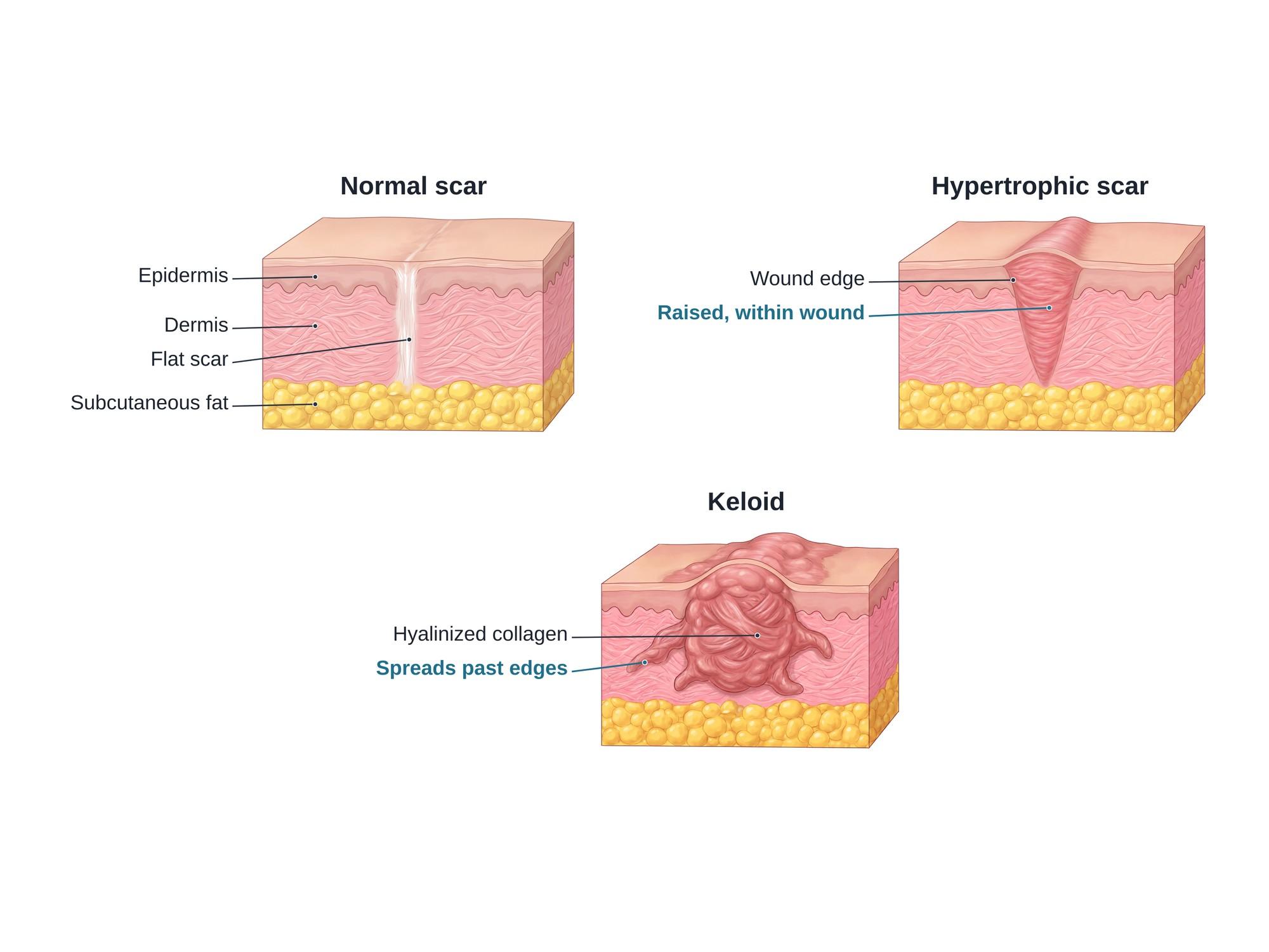
A hypertrophic scar is a thick, raised scar that forms when a wound makes too much collagen, the tough protein fiber that gives skin its strength. Skin has a thin outer layer (the epidermis), a thick collagen layer called the dermis, and fat below. The scar forms when a wound reaches the deep part of the dermis, the reticular dermis, and the new collagen piles up in thick bundles that raise the scar. It stays within the edges of the original wound. That is the key difference from a keloid, which grows past the wound edges into normal skin. Hypertrophic scars are common after burns, surgery and deep cuts. They are often red, firm and itchy. Many flatten over one to two years, but some stay thick or limit movement. Up to 70% of patients develop hypertrophic scars after burns (Finnerty et al., 2016). The figure above compares a normal flat scar, a hypertrophic scar and a keloid.
Hypertrophic scar vs keloid
The two are easy to confuse. Both are raised, firm and itchy. The difference is mostly in the borders and in how they behave over time.
| Feature | Normal scar | Hypertrophic scar | Keloid |
|---|---|---|---|
| Height | Flat, level with the skin | Raised | Raised, often much higher |
| Borders | Within the wound | Within the original wound edges | Spreads past the wound edges |
| When it appears | As the wound heals | Within weeks to months | Can appear months to years later |
| Course | Fades and softens | Often flattens over 1 to 2 years | Rarely shrinks on its own |
| Collagen under a microscope | Fine, parallel fibers | Nodules and wavy parallel bundles | Thick, glassy (hyalinized) bundles |
| After surgical removal | Not needed | Often improves | Often comes back |
Keloids are hard to treat. When a keloid is simply cut out, it usually grows back. In a meta-analysis of 608 keloids, cutting out only the inside of the keloid and leaving a thin rim cut the regrowth rate to 13% (Lawera et al., 2024). That is why doctors try to tell the two apart before planning treatment.
Causes
A hypertrophic scar can follow any injury that reaches the deep dermis. One review states that superficial injuries that do not reach the reticular dermis never cause keloids or hypertrophic scars (Ogawa, 2017).
- Burns, especially deep partial-thickness and full-thickness burns
- Surgical incisions, mainly over the chest, shoulders and joints
- Deep cuts, such as a laceration from glass or metal
- Deep scrapes, such as an abrasion or road rash from a motorcycle crash
- Skin loss, as in an avulsion injury or a degloving injury
- Infected or slow-healing wounds, and wounds that reopen
- Piercings, acne and insect bites, less often
Hypertrophic scars are common after burns. In a study of 110 burn survivors, 67% had at least one (Bombaro et al., 2003). Several things raise the risk. A systematic review of burn scarring found that these included dark skin, female sex, young age, burns on the neck and upper limb, many operations, meshed skin grafts, slow healing and more severe burns (Lawrence et al., 2012). Tension across the wound matters too. That is why scars over joints and the chest thicken more often.
Symptoms
- A raised, firm scar that stays within the wound borders
- Red or purple color while the scar is active
- Itching, often strong
- Pain or tenderness, especially when touched or stretched
- Tight skin that can limit movement over a joint
- A shiny, smooth surface with no hair or sweat
Diagnosis
Diagnosis is made by looking at the scar and asking about its history. The doctor checks its height, color, firmness and borders. The main question is whether the scar stays within the original wound or spreads past it. Scar scales such as the Vancouver Scar Scale give scores for color, height, softness and blood flow. These scores help track the scar over time.
A biopsy is rarely needed. Under a microscope, a hypertrophic scar shows nodules of collagen and fibers running in wavy bundles parallel to the skin. A keloid shows thick, glassy collagen bundles. A biopsy may be done if the diagnosis is unclear or if a skin cancer is a concern.
Treatment
Treatment aims to flatten and soften the scar, ease itching and restore movement. Most plans combine several methods.
- Silicone gel or sheets. These are worn on the scar for many hours a day, for months. In people prone to bad scars, silicone sheets cut the rate of hypertrophic scarring by about half, though the evidence is weak (O'Brien and Jones, 2013).
- Pressure garments. Tight custom garments are standard after large burns. They are worn most of the day for a year or more.
- Steroid injections. A corticosteroid such as triamcinolone is injected into the scar. It softens and flattens it over several sessions.
- Laser therapy. Pulsed dye lasers reduce redness. Fractional lasers soften thick burn scars.
- Massage and taping. These help soften scars and reduce tension on the wound.
- Surgery. Scar revision, Z-plasty or skin grafts release tight scars that limit movement. Surgery is usually combined with other treatments to stop the scar from coming back.
An international expert panel reviewed the evidence on scar care. It found "a primary role for silicone gel sheeting and intralesional corticosteroids" in managing abnormal scars (Mustoe et al., 2002).
Recovery
Scars change for a long time after the wound closes. A new scar is red and raised at first. Over months, the extra collagen slowly breaks down and lines up better. Many hypertrophic scars flatten and fade within one to two years. Burn scars can take longer.
Treatment works best when it starts early, while the scar is still active. Sun protection keeps the scar from darkening. Stretching and therapy help keep joints moving while the scar matures.
Complications
- Contracture, a tight scar that pulls the skin and limits movement, common after burns over joints
- Chronic itching and pain, which can disturb sleep
- Skin breakdown over a tight or fragile scar
- Visible disfigurement, with effects on mood, confidence and social life
- Need for more surgery, sometimes several operations over years
References
- Finnerty CC, Jeschke MG, Branski LK, Barret JP, Dziewulski P, Herndon DN. Hypertrophic scarring: the greatest unmet challenge after burn injury. Lancet. 2016.
- Ogawa R. Keloid and hypertrophic scars are the result of chronic inflammation in the reticular dermis. International Journal of Molecular Sciences. 2017.
- Lawera NG, Madzia J, Casey LC, et al. Keloid intralesional excision reduces recurrence: a meta-analytic study of the available literature on 608 keloids. Plastic and Reconstructive Surgery Global Open. 2024.
- Lawrence JW, Mason ST, Schomer K, Klein MB. Epidemiology and impact of scarring after burn injury: a systematic review of the literature. Journal of Burn Care and Research. 2012.
- Bombaro KM, Engrav LH, Carrougher GJ, et al. What is the prevalence of hypertrophic scarring following burns? Burns. 2003.
- O'Brien L, Jones DJ. Silicone gel sheeting for preventing and treating hypertrophic and keloid scars. Cochrane Database of Systematic Reviews. 2013.
- Mustoe TA, Cooter RD, Gold MH, et al. International clinical recommendations on scar management. Plastic and Reconstructive Surgery. 2002.
FAQ
Are hypertrophic scars permanent?+
Not always. Many hypertrophic scars slowly flatten and fade over one to two years as the scar matures. Some stay thick, firm or tight, especially after burns or over joints. Treatment such as silicone, pressure, steroid injections or laser can speed up and improve the result.
What is the difference between a hypertrophic scar and a keloid?+
Both are raised scars made of too much collagen. A hypertrophic scar stays within the borders of the original wound and often improves with time. A keloid grows past the wound edges into normal skin, rarely shrinks on its own, and often comes back after it is cut out.
How do you flatten a hypertrophic scar?+
Doctors usually start with silicone gel or silicone sheets and firm pressure. Steroid injections into the scar soften and flatten it. Laser treatment helps redness and thickness. Surgery is saved for scars that stay thick or tight, and is often combined with these other treatments.
Why are scars shiny?+
The skin over a scar is thin and flat. It lacks the normal ridges, hair follicles and sweat glands of healthy skin. That smooth surface reflects more light, so the scar looks shiny.
What causes a hypertrophic scar?+
Any injury that reaches the deep layer of the dermis can cause one. Common causes are burns, surgery, deep cuts, scrapes and infected wounds. Tension on the wound, slow healing and long inflammation all raise the risk.
When does a hypertrophic scar form?+
It usually appears within the first weeks to months after the wound closes. The scar turns red, raised and itchy instead of flattening. It may keep thickening for several months before it starts to settle.
Can a hypertrophic scar turn into a keloid?+
They are separate types of scar, but early on they can look alike. A raised scar that keeps growing past the edges of the wound is a keloid, not a hypertrophic scar. A doctor can usually tell them apart by the scar's borders and how it behaves over time.
Need this illustration for a patient or a case?
Describe the injury or upload a scan and get a clear, labeled medical illustration for a consult, a chart or a case exhibit.
Try Natomy →