Compression Fracture: Healing, X-ray & Life Expectancy
A compression fracture is a collapse of a vertebral body, often into a wedge. See an L4 compression fracture on X-ray beside a labeled illustration.
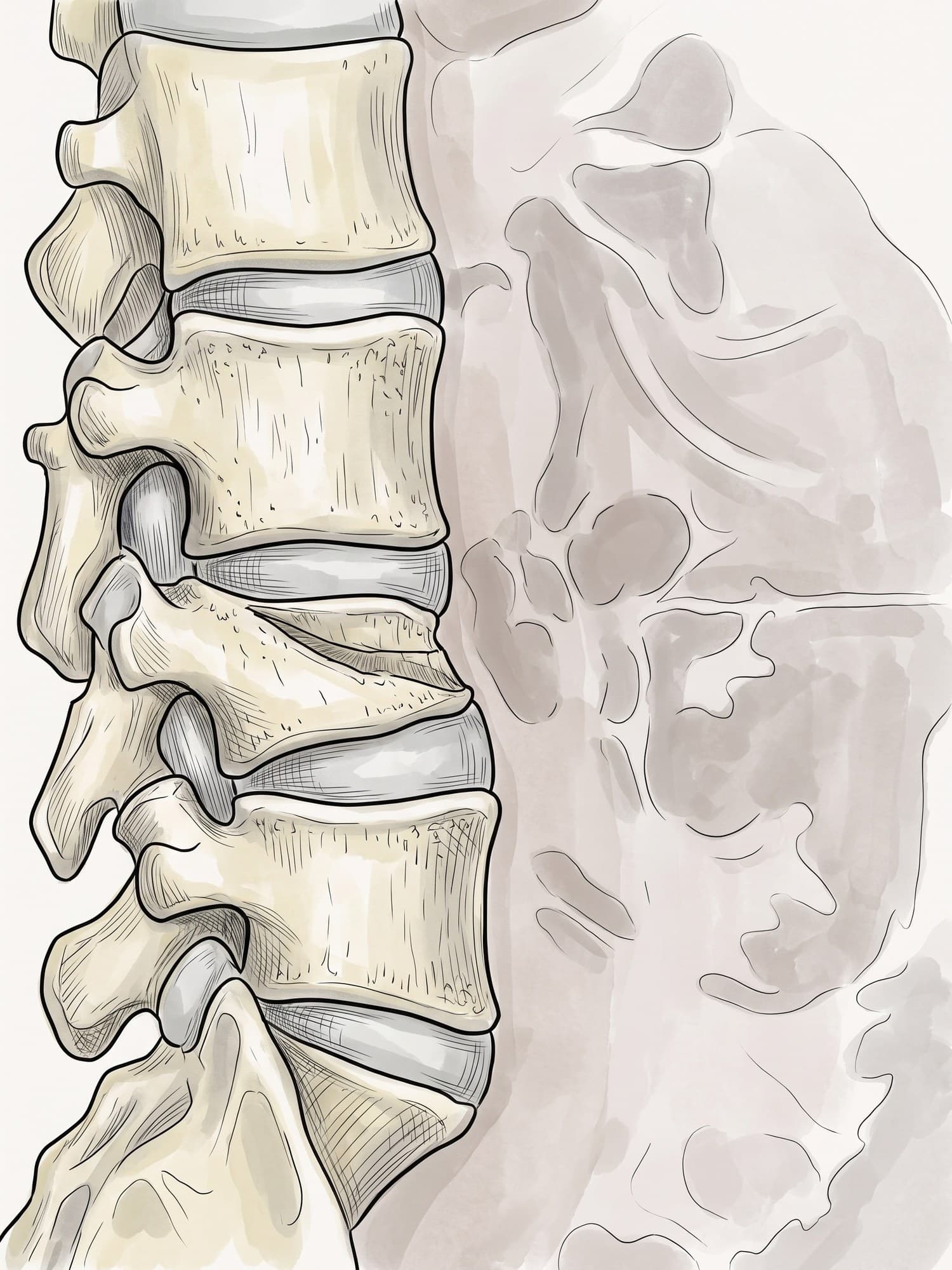
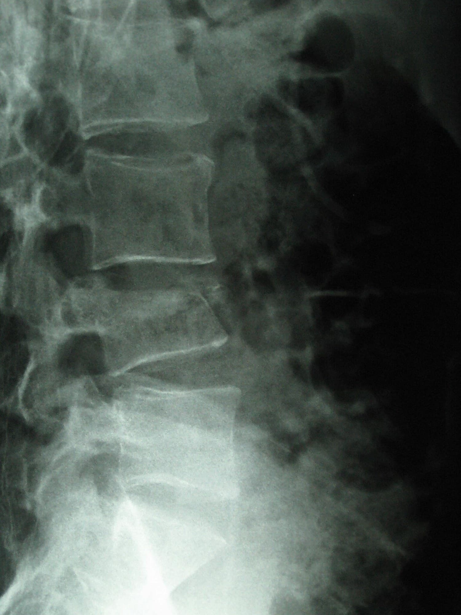
A compression fracture is a break in which the vertebral body, the solid block at the front of a vertebra, collapses under pressure. The front usually collapses more than the back, leaving the vertebra shaped like a wedge. Most compression fractures happen in the lower thoracic and upper lumbar spine, from osteoporosis or a fall. The figure above shows a real side view X-ray of an L4 compression fracture with a medical illustration drawn from it: drag the slider to compare them.
The vertebral body
Each vertebra has a cylindrical vertebral body in front, which carries the body's weight, and a ring of bone behind it that protects the spinal cord and nerves. Discs between the bodies act as cushions. In a compression fracture the vertebral body is squeezed shorter, most often at the front, while the back wall and the bony ring behind it stay intact. When the back wall breaks too and fragments push toward the spinal canal, it is a burst fracture, a more serious injury. For the normal anatomy of the lower back, see lumbar spine anatomy and thoracic spine anatomy.
Compression fracture on X-ray
The side (lateral) view shows it best. In the X-ray above, the fourth lumbar vertebra (L4) is shorter at the front than at the back, while the vertebrae above and below keep their normal rectangular shape. This patient was injured in a fall from a height.
What the radiologist describes:
| Finding | What it means |
|---|---|
| Loss of height (mild about 20 to 25%, moderate 25 to 40%, severe over 40%) | How much the body has collapsed (Genant grading) |
| Shape: wedge, biconcave or crush | Front collapse, center collapse, or whole body collapse |
| Back wall of the vertebral body | Intact in a compression fracture; broken in a burst fracture |
| Kyphosis angle | How much the spine is bent forward at the fracture |
MRI shows whether a fracture is new (bone marrow swelling) or old, which matters for treatment, and CT is used when a burst fracture is suspected.
Causes
- Osteoporosis: by far the most common cause. Weak bone can collapse from a minor fall, lifting, coughing, or with no clear injury. Vertebral fractures are the most common osteoporotic fracture.
- Trauma: falls from a height, car crashes and landing on the feet or buttocks can compress healthy vertebrae, as in the X-ray above.
- Cancer: tumors that spread to the spine, and multiple myeloma, weaken the bone and can cause collapse.
Symptoms
- Sudden back pain at the level of the fracture
- Pain worse with standing, walking, bending and lifting, and eased by lying down
- Loss of height, sometimes noticed over years
- A rounded upper back (kyphosis) after several fractures
- Rarely, numbness or weakness in the legs, which needs urgent assessment
Treatment
Most compression fractures are stable and heal without surgery.
- Pain control and activity: pain medicine and a short period of reduced activity, then walking as soon as possible to avoid deconditioning.
- Bracing: sometimes used for comfort for 6 to 12 weeks.
- Osteoporosis treatment: bone density testing and medication after any osteoporotic fracture, because one fracture greatly increases the risk of the next.
- Vertebroplasty or kyphoplasty: bone cement injected into the vertebra (in kyphoplasty, after a balloon creates a space) for fractures that remain very painful.
- Surgery with screws and rods: for unstable fractures, burst fractures, or nerve compression.
Recovery time
Most compression fractures heal in about 8 to 12 weeks, with pain easing over the first weeks. Physical therapy for posture and back extensor strength helps prevent further fractures. Some people, especially after multiple fractures, have chronic back pain.
Complications
- Chronic back pain
- Kyphosis and loss of height after multiple fractures
- Further vertebral fractures, especially in untreated osteoporosis
- Reduced lung capacity and digestive discomfort from a shortened trunk
- Progressive collapse of the fractured vertebra in the weeks after injury
How a compression fracture is shown in a personal injury case
Lumbar and thoracic compression fractures are common in rear-end and rollover collisions, falls from ladders and scaffolding, and falls in nursing homes. They are often disputed: the defense may argue the fracture was old or caused by osteoporosis, and MRI findings of bone marrow swelling become central. An illustration drawn from the plaintiff's own imaging, like the one above, shows which vertebra collapsed and by how much, and a series can show the vertebra before and after. For how these images are used at trial, see demonstrative evidence and medical legal illustration.
Related pages
FAQ
How long does a compression fracture take to heal?+
Most compression fractures heal in about 8 to 12 weeks. Pain is usually worst in the first few weeks and improves steadily, though some people have back pain for months. Healing can be slower in older adults with severe osteoporosis.
Will a compression fracture heal on its own?+
Most do. The vertebra heals in its new, flattened shape over about 3 months, with pain medicine, activity as tolerated and sometimes a brace. It does not regain its original height. Procedures such as kyphoplasty are considered when pain stays severe despite treatment.
What is the life expectancy after a compression fracture?+
A compression fracture itself is rarely life-threatening, but in older adults an osteoporotic vertebral fracture is linked with higher mortality over the following years, largely because it signals frail bones and overall poor health, and because pain can limit mobility and breathing. Treating the osteoporosis and staying active lowers the risk of further fractures.
What are the long-term effects of a spinal compression fracture?+
Some people recover with little lasting effect. Others have chronic back pain, loss of height, a forward curve of the upper back (kyphosis) when several vertebrae collapse, and reduced lung capacity or a feeling of fullness after eating as the trunk shortens. One osteoporotic fracture greatly raises the risk of another.
What does a compression fracture feel like?+
Usually a sudden, sharp back pain at the level of the fracture, worse when standing, walking, bending or twisting and eased by lying down. Some osteoporotic fractures cause only mild aching or no pain at all and are found on an X-ray taken for something else.
What is the best way to sleep with a compression fracture?+
Most people are most comfortable on their back with a pillow under the knees, or on their side with a pillow between the knees, keeping the spine straight. Getting in and out of bed by rolling onto the side and pushing up with the arms (log rolling) avoids twisting the spine.
How long do you wear a back brace for a compression fracture?+
When a brace is used, it is typically worn while up and about for about 6 to 12 weeks, mainly for comfort. Evidence that bracing speeds healing is limited, and many stable osteoporotic fractures are treated without one.
Need an illustration of a patient's own X-ray?
Upload an X-ray, CT or MRI and turn it into a clear, labeled medical illustration for a consult, a chart or a case exhibit.
Try Natomy →