Seroma vs Hematoma: Causes, Symptoms & Treatment
A seroma is a pocket of clear fluid under a surgical incision. Learn the causes, treatment, and how it differs from a hematoma.
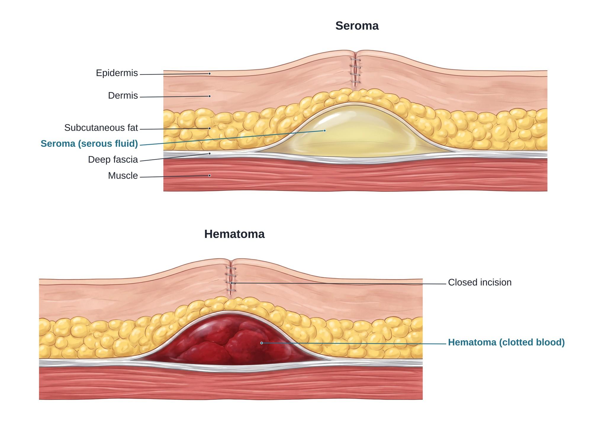
A seroma is a pocket of clear fluid that collects under the skin, usually near a surgical incision. The fluid is serous fluid, a thin, pale yellow liquid made from blood plasma and lymph. It builds up in dead space, an empty gap left when surgery separates layers of tissue. The gap usually lies between the subcutaneous fat under the skin and the deep fascia, the strong sheet that wraps the muscle. Surgery often lifts the fat off the fascia as a flap and cuts small lymph and blood vessels. If the flap does not stick back down, fluid from them fills the gap. Seroma is one of the most common complications after surgery. After a tummy tuck (abdominoplasty), about 1 in 5 patients (19.5%) developed a seroma when no extra prevention steps were used. With prevention steps, the rate fell to 7.5% (Seretis et al., 2017). Most seromas go away on their own. The figure above shows a seroma beside a hematoma in the same layer.
Seroma vs hematoma
A seroma and a hematoma can sit in the same place, as the figure shows. What fills the pocket is different.
| Feature | Seroma | Hematoma |
|---|---|---|
| Contents | Clear, thin, pale yellow serous fluid | Blood, often clotted |
| Usual onset | Days to weeks after surgery | Hours to a few days after surgery |
| How it feels | Soft, fluid moves when pressed | Firmer, may feel lumpy |
| Skin color | Usually normal | Often bruised, purple or blue |
| Usual cause | Leaking lymph and plasma into dead space | Bleeding from a vessel |
| Usual treatment | Wait, or drain with a needle | Wait, or open and remove the clot |
A hematoma can turn into a seroma over time. As the clot breaks down, its liquid part can stay behind as a clear or brown-tinged collection. Blood that spreads into the tissue instead of pooling is a bruise, or contusion.
Types
Seromas have no single grading system. In hernia surgery, one widely used scheme groups them by how long they last and what treatment they need (Morales-Conde, 2012).
| Type | What it means |
|---|---|
| Type 0 | No seroma on exam (0a), or seen only on imaging (0b) |
| Type I | Seroma on exam that lasts less than 1 month |
| Type II | Seroma that lasts 1 to 6 months |
| Type III | Seroma with minor problems, such as pain, a lasting bulge or superficial infection, that may need medical treatment |
| Type IV | Seroma that must be treated, such as drained by needle or surgery, or one that leaks or becomes deeply infected |
Doctors also describe a seroma as acute (new) or chronic. A chronic seroma can grow a smooth wall of scar tissue around it. This wall is called a pseudocapsule or pseudobursa. It keeps making fluid, so the pocket keeps refilling.
Causes
Any operation that leaves dead space can cause a seroma. Common ones include:
- Breast surgery, especially mastectomy and removal of armpit lymph nodes (axillary dissection)
- Abdominoplasty (tummy tuck) and liposuction
- Hernia repair, especially with mesh
- Cesarean section and other abdominal surgery
- Removal of large lumps or masses
- Flap surgery, where tissue is moved to rebuild another area
Some factors raise the risk. Being overweight is one of the clearest. After a tummy tuck, 38% of overweight or obese patients developed a seroma, compared with 19% of patients at normal weight (Kim and Stevenson, 2006). The type of operation also matters. In one study of 158 breast cancer patients, 35% developed a seroma, and the risk was higher after modified radical mastectomy (Hashemi et al., 2004). Other factors include large areas of dissection, cutting with electrocautery, removing drains too early, and moving the area too much too soon.
Seromas also form after injuries, not just surgery. A hard blow or a shearing force can tear the fat away from the fascia without breaking the skin. Fluid then fills the gap. This is called a Morel-Lavallée lesion, a closed form of degloving injury. It is common over the hip and thigh after car and motorcycle crashes.
Symptoms
- A soft, smooth swelling under or near the incision
- A feeling of fluid moving when the area is pressed (fluctuance)
- Tightness, pressure or mild pain
- Clear or pale yellow fluid leaking from the incision
- Swelling that grows over days rather than going down
Warning signs of infection include redness, warmth, fever, worsening pain, or cloudy or foul-smelling drainage.
Diagnosis
A seroma is usually found on physical exam. The doctor feels a soft, fluid-filled swelling near the wound. An ultrasound can confirm it. On ultrasound, a seroma looks like a dark, fluid-filled space with smooth edges. A hematoma tends to look more mixed, because clots reflect sound in a patchy way.
If the doctor drains the pocket, the fluid itself helps tell them apart. Seroma fluid is clear and straw-colored. Hematoma fluid is dark red, and clots may not come out through a needle. Cloudy fluid suggests infection and is sent to the lab.
Treatment
- Watchful waiting. Small seromas with no symptoms are often left alone. The body absorbs the fluid over weeks.
- Needle aspiration. The doctor numbs the skin and drains the fluid with a sterile needle and syringe. This may be repeated if the fluid comes back.
- Drains. A thin tube can be placed to let fluid flow out for days or weeks.
- Compression. A binder or compression garment may be used to help the layers stick together.
- Sclerotherapy. A substance is injected into the pocket to irritate its walls so they scar together.
- Surgery. A chronic seroma with a thick capsule may need to be opened, the capsule removed, and the space closed.
Prevention starts in the operating room. A systematic review of 75 studies with 7,173 patients found several methods that lowered seroma risk (Janis et al., 2016). These included closed-suction drains kept in until output was low, quilting or progressive tension sutures (stitches that tack the flap down to the fascia to close dead space), sharp or ultrasonic dissection instead of cautery, and rest for the surgical area. Compression alone did not prevent seromas in that review.
Recovery
Most seromas resolve within a few weeks to a few months. Some need several drainages before they stop refilling. Recovery is slower when the seroma is large, keeps coming back, or becomes infected. A seroma can also delay the next step of care, such as chemotherapy or radiation after cancer surgery.
Complications
- Infection, which can turn the seroma into an abscess
- Wound dehiscence, where pressure from the fluid pulls the incision apart
- Skin flap necrosis, death of thin skin over a large pocket
- Chronic seroma with a pseudocapsule that keeps refilling
- Visible bulge or contour deformity, especially after cosmetic surgery
- Scarring, including raised hypertrophic scars at an incision that healed under tension
References
- Seretis K, Goulis D, Demiri EC, Lykoudis EG. Prevention of seroma formation following abdominoplasty: a systematic review and meta-analysis. Aesthetic Surgery Journal. 2017.
- Morales-Conde S. A new classification for seroma after laparoscopic ventral hernia repair. Hernia. 2012.
- Kim J, Stevenson TR. Abdominoplasty, liposuction of the flanks, and obesity: analyzing risk factors for seroma formation. Plastic and Reconstructive Surgery. 2006.
- Hashemi E, Kaviani A, Najafi M, et al. Seroma formation after surgery for breast cancer. World Journal of Surgical Oncology. 2004.
- Janis JE, Khansa L, Khansa I. Strategies for postoperative seroma prevention: a systematic review. Plastic and Reconstructive Surgery. 2016.
FAQ
When does a seroma go away?+
Most small seromas are absorbed by the body over a few weeks to a few months. Larger ones may need to be drained with a needle once or several times. A seroma that lasts for months can form a scar-like capsule and may need surgery.
How do you help a seroma reabsorb?+
Follow the surgeon's advice. This often means wearing a compression garment or binder, keeping drains in until the output is low, and limiting movement of the area for a short time. Do not try to drain or squeeze it yourself. Large or painful seromas are drained by a doctor with a sterile needle.
What is the difference between a seroma and a hematoma?+
Both are pockets of fluid under a wound. A seroma holds clear, thin, pale yellow fluid made from plasma and lymph. A hematoma holds blood, which often clots. A hematoma usually appears sooner, feels firmer, and may bruise the skin. A seroma feels soft and moves like a water balloon.
Is a seroma dangerous?+
Most seromas are not dangerous. They are uncomfortable and slow down recovery. The main risks are infection, pressure on the incision that pulls it open, and a chronic seroma with a capsule. Redness, warmth, fever or cloudy drainage need prompt care.
Can a seroma burst?+
A seroma can leak through the incision, often as a sudden gush of clear or pink-tinged fluid. This is not usually an emergency, but the surgeon should check the wound. Leaking through the incision can delay healing and raise the risk of infection.
How many times can a seroma be drained?+
There is no fixed limit. Many seromas need two or three needle drainages before they stop refilling. If one keeps coming back, the surgeon may place a drain, inject a sclerosing agent, or remove the capsule in surgery.
Is it normal to have a seroma after a lumpectomy or mastectomy?+
Yes. Seroma is the most common complication after breast cancer surgery, especially when lymph nodes in the armpit are removed. Most are small and go away with time or a few drainages.
What is considered a large seroma?+
There is no single cutoff. Surgeons usually call a seroma large when it is visible as a bulge, causes pain or tightness, stretches the incision, or needs to be drained. Some hold more than a cup of fluid.
Need this illustration for a patient or a case?
Describe the injury or upload a scan and get a clear, labeled medical illustration for a consult, a chart or a case exhibit.
Try Natomy →