Retinal Detachment: Symptoms, Causes & Treatment
Retinal detachment is when the retina lifts off the back of the eye, often after a tear. Learn the types, symptoms, causes and treatment.
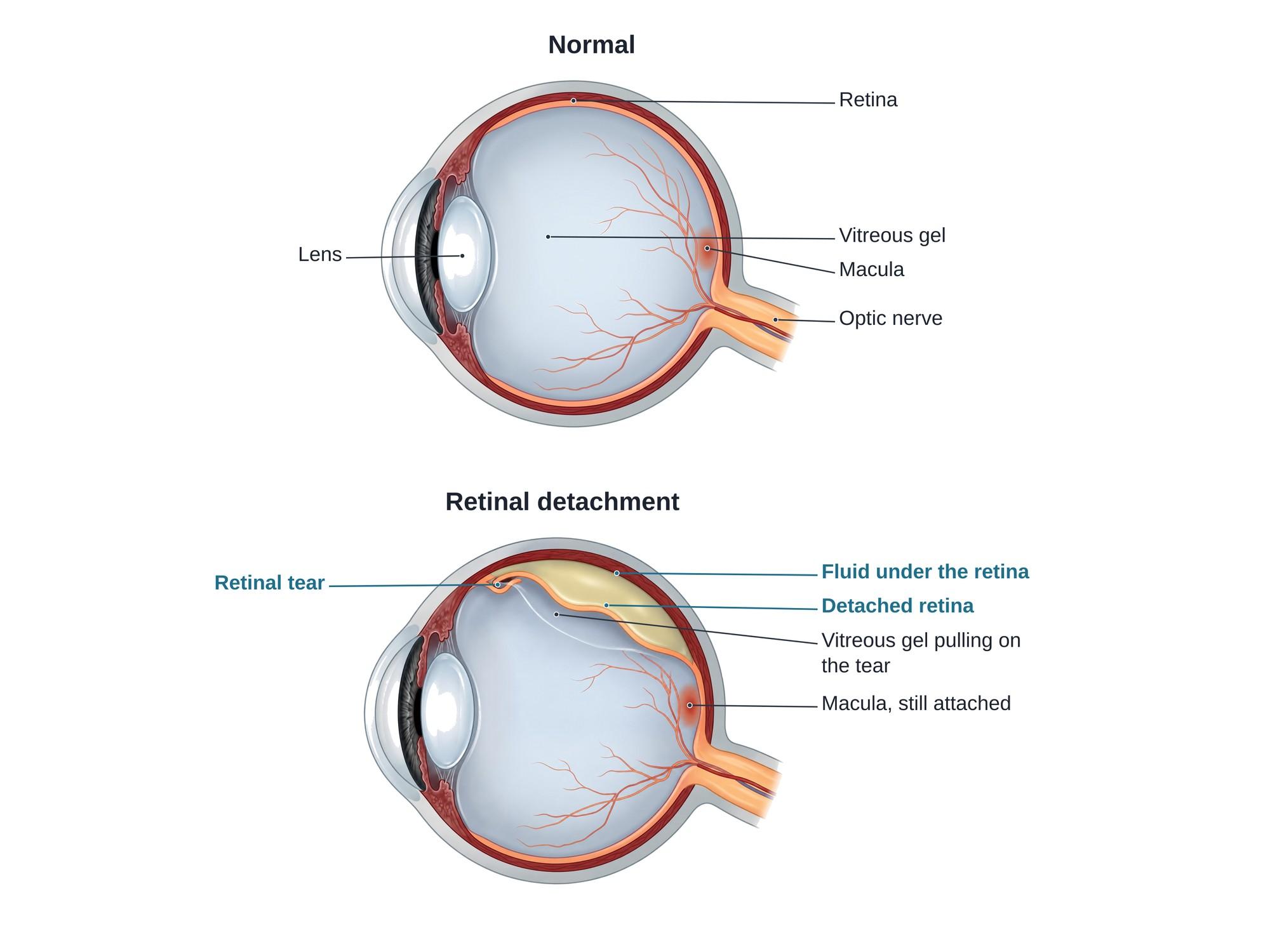
A retinal detachment is when the retina, the thin light-sensing layer at the back of the eye, lifts off the wall of the eye. Once it is detached, the retina loses its blood supply and stops working. The most common type is a rhegmatogenous retinal detachment, which starts with a tear in the retina. Fluid passes through the tear and collects under the retina, lifting it further. About 1 in 10,000 people develop one each year (Ge et al., 2024). It is painless, but it is an emergency, because a detachment left untreated can cause permanent vision loss. The illustration above shows a normal eye beside an eye with a rhegmatogenous detachment.
The anatomy
The wall of the eye has three main layers. The tough white outer coat is the sclera. Inside it is the choroid, a layer rich in blood vessels. The innermost layer is the retina.
The retina has two parts. The neurosensory retina holds the light-sensing cells (photoreceptors). Under it lies a single layer of pigment cells, the retinal pigment epithelium (RPE), which sticks to the choroid. The two parts are not firmly glued together. A retinal detachment is a split between them.
The large space in front of the retina is filled with the vitreous, a clear gel. The gel is stuck to the retina in some places, most firmly at its front edge.
Types
| Type | What causes it | Key feature |
|---|---|---|
| Rhegmatogenous | A tear or hole in the retina | Fluid passes through the break and lifts the retina. The most common type. |
| Tractional | Scar tissue on the retina contracts and pulls it off | No tear. Often from diabetic eye disease. |
| Exudative (serous) | Fluid leaks from under the retina | No tear. Caused by inflammation, tumors or other eye disease. |
The macula is the center of the retina. It gives sharp central vision for reading and faces. Doctors also describe a detachment as macula-on (the macula is still attached, as in the illustration) or macula-off (the macula has detached). Macula-on detachments usually keep better central vision after surgery.
Causes
Most detachments start with a posterior vitreous detachment (PVD). With age, the vitreous gel shrinks and peels away from the retina. Where the gel is stuck firmly, it can pull a flap out of the retina, a horseshoe tear. Watery fluid then flows through the tear and under the retina. In a review of 17 studies, 14% of patients seen by an eye doctor for new floaters or flashes had a retinal tear (Hollands et al., 2009).
Risk factors include:
- Older age, as the vitreous gel shrinks and separates
- Nearsightedness (myopia), which stretches the retina and makes it thinner
- Past cataract surgery: about 1 in 5 rhegmatogenous detachments in the UK occur in eyes that have had it (Sultan et al., 2020)
- Lattice degeneration, thin weak patches in the outer retina
- A detachment in the other eye: in a Scottish study, 7% of patients had detachments in both eyes (Sultan et al., 2020)
- Eye trauma, such as a punch, a ball, a fall, an airbag or a penetrating injury
- Family history of retinal detachment
Symptoms
- Flashes of light (photopsia), often at the edge of vision
- A sudden shower of new floaters, such as dots, threads or cobwebs
- A dark shadow or curtain moving across part of the vision
- Blurred or distorted vision, especially once the macula is involved
Flashes, floaters and field loss are the "three F's." A detachment does not hurt, so people sometimes wait. Anyone with these symptoms needs an eye exam the same day.
Diagnosis
An eye doctor dilates the pupils with drops and looks at the whole retina.
- Dilated fundus exam: the doctor sees the lifted gray retina, the tear and how far the detachment reaches. Gently pressing on the eyelid (scleral depression) shows the far edge of the retina.
- Slit lamp exam: pigment cells floating in the vitreous ("tobacco dust") suggest a tear.
- Optical coherence tomography (OCT): a light scan that shows whether the macula is still attached.
- Ultrasound of the eye: shows the detached retina as a raised membrane when blood or a cloudy lens blocks the view.
Treatment
A retinal tear without a detachment is sealed with laser or cryotherapy (freezing), usually in the office. This creates a scar that welds the retina around the tear.
A retinal detachment needs surgery to close the tear and put the retina back.
- Pneumatic retinopexy: a gas bubble is injected into the eye. The patient positions their head so the bubble presses the retina back over the tear, which is then sealed with laser or freezing.
- Scleral buckle: a silicone band is sewn onto the outside of the eye. It pushes the wall in toward the tear and relieves the pull of the gel.
- Vitrectomy: the surgeon removes the vitreous gel, flattens the retina, seals the tear with laser and fills the eye with gas or silicone oil.
In a randomized trial of 176 patients with suitable detachments, one surgery reattached the retina in 81% after pneumatic retinopexy and 93% after vitrectomy. With further surgery, more than 98% in both groups were reattached (Hillier et al., 2019). The same trial found better vision at one year after pneumatic retinopexy.
Recovery
Vision recovers slowly over weeks to months. If gas was used, the patient may need to hold a set head position for several days. They must not fly or go to high altitude until the gas is gone, because the bubble expands. Silicone oil usually needs a second operation to remove it. After a vitrectomy, a cataract often forms within a year or two. When the macula was detached, central vision is often less sharp than before, even after a successful repair.
Complications
- Redetachment, often from new tears or scar tissue
- Proliferative vitreoretinopathy (PVR), scar tissue that grows on the retina and pulls it off again
- Permanent vision loss, especially after a long macula-off detachment
- Distorted vision (metamorphopsia) or double vision
- Cataract after vitrectomy
- Raised eye pressure from gas or silicone oil
- Detachment in the other eye
How retinal detachment is shown in a personal injury case
Retinal detachment follows blunt eye injuries from assaults, falls, sports, workplace accidents and car crashes, including airbag strikes. Trauma can also cause bleeding in the front of the eye (hyphema), which can hide the retina from view at first. A traumatic detachment can show up weeks, months or even years after the injury. Medical records may show a retinal dialysis, a giant tear or a vitreous hemorrhage. A clear illustration helps a jury see where the tear formed, how fluid lifted the retina, and why the macula's status mattered for vision. For how these images are used at trial, see demonstrative evidence and medical legal illustration.
References
- Ge JY, Teo ZL, Chee ML, et al. International incidence and temporal trends for rhegmatogenous retinal detachment: a systematic review and meta-analysis. Survey of Ophthalmology. 2024;69(3):330-336.
- Hollands H, Johnson D, Brox AC, et al. Acute-onset floaters and flashes: is this patient at risk for retinal detachment? JAMA. 2009;302(20):2243-2249.
- Sultan ZN, Agorogiannis EI, Iannetta D, et al. Rhegmatogenous retinal detachment: a review of current practice in diagnosis and management. BMJ Open Ophthalmology. 2020;5(1):e000474.
- Hillier RJ, Felfeli T, Berger AR, et al. The Pneumatic Retinopexy versus Vitrectomy for the Management of Primary Rhegmatogenous Retinal Detachment Outcomes Randomized Trial (PIVOT). Ophthalmology. 2019;126(4):531-539.
FAQ
Can a torn retina cause blindness?+
Yes, if it is not treated. A tear lets fluid get under the retina and lift it off. A detached retina stops working, and if it is left detached, vision in that eye can be lost for good. Most tears found early can be sealed with laser or freezing treatment in the office, which usually prevents a detachment.
What do they do for a detached retina?+
Surgery reattaches the retina and seals the tear. The main options are pneumatic retinopexy (a gas bubble injected into the eye), a scleral buckle (a band sewn around the outside of the eye), and vitrectomy (removing the vitreous gel and filling the eye with gas or silicone oil). The choice depends on where the tear is, how large the detachment is, and the eye's history.
What are the first signs of a detached retina?+
New flashes of light, a sudden shower of floaters, and then a dark shadow or curtain spreading across part of the vision. Some people only notice blurred vision. The detachment itself is painless. These symptoms need an eye exam the same day.
How long before retinal detachment causes blindness?+
There is no fixed time. Some detachments spread over hours, others over days or weeks. Once the macula detaches, central vision drops sharply, and the longer it stays off, the less vision usually returns. That is why a detachment that has not yet reached the macula is treated as an emergency.
Can a blow to the eye cause retinal detachment?+
Yes. Blunt trauma, such as a punch, a ball, a fall or an airbag, can tear the retina or pull it away from its front edge (a retinal dialysis). The detachment may appear soon after the injury or months to years later, so injured eyes need a dilated exam and follow-up.
Can lack of sleep or stress cause retinal detachment?+
No. Neither lack of sleep nor stress is a known cause. The main causes are age-related changes in the vitreous gel, nearsightedness, past eye surgery such as cataract surgery, and eye injury.
How can I avoid retinal detachment?+
Most detachments cannot be fully prevented, but the risk can be lowered. Wear eye protection for sports and risky work. Know the warning signs and get seen the same day if they appear. People with high myopia, lattice degeneration, past cataract surgery or a detachment in the other eye should have regular dilated eye exams.
How long does it take to recover from retinal detachment surgery?+
The eye usually takes several weeks to settle, and vision can keep improving for months. If a gas bubble was used, the patient may need to hold a set head position for days and must not fly or travel to high altitude until the gas is gone. Vision is often less sharp than before if the macula was detached.
Need this illustration for a patient or a case?
Describe the injury or upload a scan and get a clear, labeled medical illustration for a consult, a chart or a case exhibit.
Try Natomy →