Hyphema: Blood in the Eye, Causes, Grades & Treatment
A hyphema is blood pooling in the front chamber of the eye, usually after a blow to the eye. See it illustrated from the side and the front.
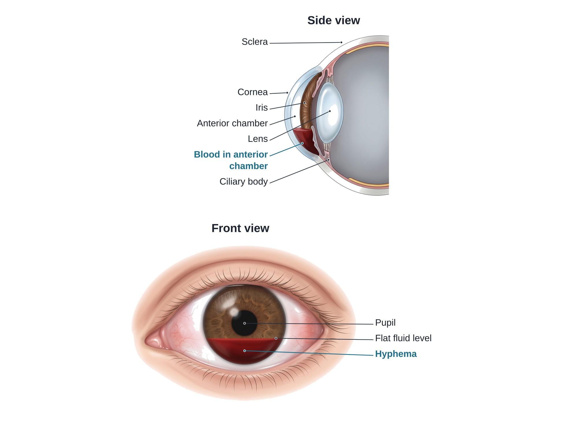
A hyphema is blood in the front chamber of the eye, the clear space between the cornea and the iris. It is usually caused by a blunt blow to the eye, such as a ball, a fist, an airbag or a fall. The blood comes from small torn vessels in the iris or at the root of the iris. Because blood is heavier than the fluid in the eye, it sinks and forms a red layer with a flat top. Most hyphemas clear in about a week, but some cause high eye pressure, rebleeding or lasting damage. The illustration above shows a hyphema from the side and from the front.
The anatomy
The front of the eye has a small fluid-filled space called the anterior chamber.
- The cornea is the clear dome at the front of the eye. It forms the front wall of the chamber.
- The iris is the colored ring behind the cornea. It forms the back wall. The pupil is the opening in its center.
- The chamber is filled with aqueous humor, a clear fluid made by the ciliary body behind the iris.
- The fluid drains out through the drainage angle, where the cornea meets the iris. The drain is a sieve-like tissue called the trabecular meshwork.
- The lens sits just behind the pupil, and the white outer wall of the eye is the sclera.
In a hyphema, blood fills part of the anterior chamber. It can block vision and clog the drain, which raises the pressure inside the eye.
Grades
Doctors grade a hyphema by how much of the anterior chamber is filled with blood.
| Grade | How much blood | What it looks like |
|---|---|---|
| Microhyphema | Red cells floating in the fluid | No layer; seen only with a slit lamp |
| Grade I | Less than one third of the chamber | A thin layer at the bottom, like the one illustrated |
| Grade II | One third to one half | The layer reaches toward the pupil |
| Grade III | Half to nearly all | Most of the iris and pupil are covered |
| Grade IV | The whole chamber | Called a total or "eight-ball" hyphema when the blood is dark and clotted |
Higher grades carry a higher risk of high eye pressure and poor vision.
Causes
- Blunt eye trauma. This is the most common cause. Sports balls, fists, elbows, paintballs, bungee cords and car airbags are typical.
- Penetrating injury, when an object cuts into the eye.
- Eye surgery, such as cataract or glaucoma surgery.
- Spontaneous hyphema, without injury. It can happen with abnormal new blood vessels on the iris (from diabetes or a blocked retinal vein), eye tumors, bleeding disorders, or blood-thinning medicines.
In blunt trauma, the force pushes the front of the eye backward. The eye stretches sideways, which tears small vessels at the root of the iris and in the ciliary body.
Symptoms
- A red or dark layer at the bottom of the colored part of the eye
- Blurred, cloudy or blocked vision
- Eye pain
- Sensitivity to light (photophobia)
- Nausea, vomiting or headache, which can mean the eye pressure is high
- A history of a recent blow to the eye
Diagnosis
An eye doctor examines the eye with a slit lamp, a microscope with a bright beam of light. The exam shows the height of the blood layer and whether there is a clot. The doctor measures the intraocular pressure (the pressure inside the eye) and checks vision.
The doctor also looks for other injuries. These include a torn iris, a dislocated lens, bleeding in the back of the eye, a detached retina, or a ruptured eyeball. If the eye could be ruptured, it is protected and not pressed on. A CT scan of the orbits may be used to look for a broken orbit bone or a foreign object. An ultrasound of the eye may be done gently if the back of the eye cannot be seen.
People of African or Mediterranean descent are often tested for sickle cell disease or trait. Sickled blood cells clog the drain more easily, and the optic nerve is more easily damaged by high pressure.
Treatment
Most hyphemas are treated without surgery. The goals are to stop rebleeding, control the pressure and protect the eye.
- Rest and a raised head. Quiet activity, with the head of the bed raised about 30 to 45 degrees.
- A rigid eye shield over the eye to prevent another injury.
- Eye drops. Steroid drops calm inflammation. Dilating drops (cycloplegics) relax the iris and ease pain.
- Pressure-lowering drops or pills when the eye pressure is high.
- Avoiding aspirin and anti-inflammatory medicines, which raise the risk of rebleeding.
- Daily or frequent checks in the first days, when rebleeding is most likely.
Surgery is used when the pressure stays high despite medicine, when blood begins to stain the cornea, or when a large clot does not clear. The surgeon washes the blood out of the anterior chamber (an anterior chamber washout). A glaucoma procedure may be added if the pressure stays high.
Recovery
- Small hyphemas often clear in about 5 to 7 days.
- Rebleeding is most likely in the first 3 to 5 days. A second bleed is often larger than the first.
- Activity, sports and heavy lifting are limited until the doctor says the eye has healed.
- Vision usually returns once the blood is gone, unless other parts of the eye were injured.
- A follow-up exam weeks later checks the drainage angle. A blow strong enough to cause a hyphema can damage the angle and lead to glaucoma years later, so yearly eye pressure checks are often advised.
Complications
- Rebleeding, which raises the risk of every other complication.
- High eye pressure from blood clogging the drain. Over time this can damage the optic nerve.
- Corneal blood staining, when blood breaks down and stains the cornea brown. It can take months to clear and can cause amblyopia (a "lazy eye") in young children.
- Angle recession glaucoma, a long-term pressure problem from a torn drainage angle.
- Synechiae, scar tissue that sticks the iris to the cornea or lens.
- Optic nerve damage and vision loss, especially in people with sickle cell disease or trait.
How hyphema is shown in a personal injury case
Hyphema is common after airbag injuries, assaults, sports injuries and workplace accidents. In a claim, the questions are often how hard the eye was hit, whether the eye was protected, and whether there is lasting damage such as glaucoma or vision loss. A clear medical illustration shows a jury where the blood collected inside the eye, why it blocked vision, and how it can clog the eye's drain and raise the pressure. Pairing a side section with a front view helps jurors match the anatomy to what the treating doctor saw. For how these images are used at trial, see demonstrative evidence and medical legal illustration.
FAQ
What are the symptoms of hyphema in the eye?+
Blurred or blocked vision, eye pain, and sensitivity to light are common. A large hyphema can be seen as a red layer at the bottom of the colored part of the eye. A very small one (a microhyphema) may only be seen by an eye doctor with a slit lamp. Pain and nausea can mean the pressure in the eye has gone up.
What is the difference between hyphema and subconjunctival hemorrhage?+
A subconjunctival hemorrhage is blood under the thin clear skin over the white of the eye. It looks like a bright red patch on the white part, does not affect vision, and is usually harmless. A hyphema is blood inside the eye, in front of the iris. It sits over the colored part of the eye, can blur vision, and needs an eye exam the same day.
What could be causing blood in the white of my eye?+
Blood on the white of the eye is usually a subconjunctival hemorrhage. It can follow coughing, straining, rubbing the eye, minor injury or blood thinners, and it usually clears in 1 to 2 weeks. Blood over the colored part of the eye, or any blood with pain or vision loss, is different and should be checked by an eye doctor right away.
Which medication should you avoid with a hyphema?+
Aspirin and anti-inflammatory pain medicines such as ibuprofen and naproxen are usually avoided, because they can raise the risk of rebleeding. Acetaminophen is often used for pain instead. Anyone on blood thinners should tell the eye doctor, but should not stop them without medical advice.
How long does a hyphema take to heal?+
A small hyphema is often absorbed in about 5 to 7 days. Larger ones take longer. The first few days matter most, because a second bleed is most likely in the first 3 to 5 days after the injury.
Can a hyphema cause blindness?+
Most hyphemas heal without lasting vision loss. Permanent damage can happen when the eye pressure stays high, when blood stains the cornea, or when the injury also damaged the lens, retina or optic nerve. People with sickle cell disease or trait are at higher risk of optic nerve damage.
Is a hyphema an emergency?+
Yes. Any visible blood in the front of the eye after an injury should be seen by an eye doctor or emergency department the same day. The doctor checks the eye pressure and looks for other injuries inside the eye.
How should you sleep with a hyphema?+
Doctors often advise sleeping with the head raised about 30 to 45 degrees. This helps the blood settle to the bottom of the chamber, away from the pupil, and can help vision clear sooner. A rigid eye shield is often worn, especially at night.
Need this illustration for a patient or a case?
Describe the injury or upload a scan and get a clear, labeled medical illustration for a consult, a chart or a case exhibit.
Try Natomy →