Photopsia (Flashes in the Eye): Causes & Warning Signs
Photopsia is seeing flashes of light, most often from the vitreous gel pulling on the retina. Learn the warning signs, and see it illustrated beside a normal eye.
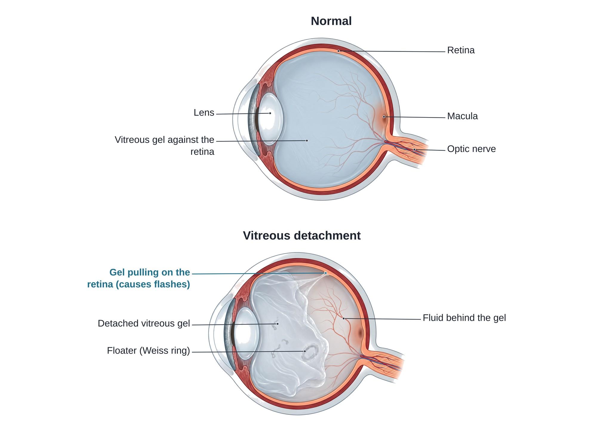
Photopsia means seeing flashes of light that are not really there. The flashes look like sparks, streaks or lightning, often at the edge of vision. The most common cause is the vitreous gel inside the eye pulling on the retina as it separates with age, called a posterior vitreous detachment. The retina reacts to the tug as if it were light. New flashes can also be a warning sign of a retinal tear or retinal detachment, which can cause blindness without prompt treatment. The illustration above shows a normal eye beside an eye with a vitreous detachment pulling on the retina.
The anatomy
The retina is the thin, light-sensing layer that lines the back of the eye. It sends signals to the brain through the optic nerve. The center of the retina, the macula, gives sharp central vision.
The large space in front of the retina is filled with the vitreous, a clear gel made mostly of water and fine collagen fibers. In young people, the gel fills the eye and lies firmly against the retina. Its outer layer, the posterior hyaloid, is stuck to the retina in some places, most strongly at the front edge (the vitreous base), around the optic nerve and along some blood vessels.
The retina can only send one kind of signal: light. So when something pulls on it or presses on it, the brain sees a flash.
How photopsia happens
With age, the vitreous gel slowly turns more watery and shrinks. At some point, often between ages 50 and 70, the gel peels away from the back of the eye. This is a posterior vitreous detachment (PVD).
- The gel collapses forward and pulls away from the retina.
- Watery fluid fills the space behind it.
- Where the gel is still stuck, it tugs on the retina. Each tug, often with eye movement, causes a flash.
- The gel's outer layer often carries a ring of tissue from around the optic nerve, called a Weiss ring. It casts a shadow on the retina and is seen as a floater.
Usually the gel lets go cleanly and the flashes stop. In some people, the gel pulls hard enough to tear the retina. Fluid can then pass through the tear and lift the retina off the back of the eye, a retinal detachment.
Types
| Type | Cause | What the flashes are like |
|---|---|---|
| Vitreoretinal (from the eye) | Vitreous detachment, retinal tear or detachment | Brief sparks or arcs in one eye, often at the side, worse in dim light and with eye movement |
| Migraine aura | Spreading change in the brain's visual cortex | Zigzag, shimmering lines that spread over 5 to 60 minutes, seen with both eyes |
| Traumatic | A blow to the eye or head | Sudden flashes or "seeing stars" at the time of injury, sometimes followed by a later retinal tear |
| Other | Eye inflammation, optic neuritis, low blood pressure, some medicines, brain conditions | Varies |
A rare type of migraine, hemiplegic migraine, can cause visual aura together with weakness on one side of the body.
Causes
- Posterior vitreous detachment, the most common cause
- Retinal tear or retinal detachment
- Migraine with aura
- Eye or head trauma, such as a punch, a ball to the eye, an airbag or a fall
- Nearsightedness (myopia), past eye surgery such as cataract surgery, and diabetes, which make vitreous and retinal problems more likely
- Inflammation inside the eye (uveitis) or of the optic nerve
- Problems in the brain's vision areas, such as stroke or a mass, which are rare
Symptoms
- Flashes of light, sparks or streaks, often at the edge of vision
- New floaters, such as specks, threads, a ring or cobwebs
- A shower of many small dots, which can mean bleeding inside the eye from a retinal tear
- A shadow or curtain moving across part of the vision, a sign of retinal detachment
- Blurred vision
Flashes, floaters and field loss are the "three F's" of a retinal tear or detachment. They need an eye exam the same day.
Diagnosis
An eye doctor dilates the pupils with drops and examines the vitreous and the whole retina, out to its far edge.
- Dilated fundus exam with an ophthalmoscope and a special lens, sometimes pressing gently on the eyelid (scleral depression) to see the far edge of the retina
- Slit lamp exam, looking for pigment cells floating in the vitreous ("tobacco dust"), a sign of a retinal tear
- Optical coherence tomography (OCT), a scan that shows whether the vitreous has separated and whether it is pulling on the macula
- Ultrasound of the eye when bleeding blocks the view
If the first exam is normal, a repeat exam is often done within a few weeks, because a tear can still form while the gel is separating.
Treatment
- Vitreous detachment without a tear: no treatment is needed. Flashes usually fade over weeks to months. The patient is told to return at once if flashes or floaters get worse or a shadow appears.
- Retinal tear: sealed with laser photocoagulation or cryotherapy (freezing), usually in the office, to stop a detachment.
- Retinal detachment: surgery, such as pneumatic retinopexy (a gas bubble), scleral buckle or vitrectomy (removing the vitreous gel).
- Migraine aura: migraine treatment and prevention.
Recovery
Flashes from a simple vitreous detachment often settle within 1 to 3 months. Floaters can last longer but usually become less noticeable. After laser for a retinal tear, most people return to normal activity within a day or two. After retinal detachment surgery, recovery takes weeks to months, and some people must keep their head in a set position while a gas bubble works.
Complications
- Retinal tear, found in a meaningful minority of people with a new, symptomatic vitreous detachment
- Retinal detachment, which can cause permanent vision loss if not treated quickly
- Vitreous hemorrhage, bleeding into the gel that blurs vision
- Macular hole or vitreomacular traction, when the gel pulls on the center of the retina
How photopsia is shown in a personal injury case
Photopsia comes up in two kinds of cases. In personal injury cases, a blow to the eye or head in a car crash, assault or fall can trigger a vitreous detachment or tear the retina, and flashes may be the first sign. In medical malpractice cases, the question is often whether a doctor, urgent care or emergency room missed the warning signs. A patient who reports new flashes and floaters needs a dilated retina exam. If that exam is delayed and the retina detaches, the person can lose vision that early laser treatment could have saved. A clear medical illustration of a normal eye beside an eye with the vitreous pulling on the retina shows a jury what causes the flashes and why they are a warning sign. For how these images are used at trial, see demonstrative evidence and medical legal illustration.
FAQ
What could be causing the small flashes of light in my vision?+
The most common cause is a posterior vitreous detachment, when the gel inside the eye pulls away from the retina with age. Other causes are a retinal tear or detachment, migraine aura, a blow to the eye or head, and, less often, eye inflammation or problems in the brain's vision areas. New flashes need an eye exam the same day or the next day.
What are the three F's of retinal detachment?+
Flashes, floaters and field loss. Flashes are brief streaks or sparks of light. Floaters are new specks, cobwebs or a sudden shower of dots. Field loss is a dark shadow or curtain spreading across part of your vision. Any of these, and especially all three together, is an emergency.
Is photopsia dangerous?+
Often not, but it can be. Most flashes come from a harmless vitreous detachment. In some people the pulling gel tears the retina, and a tear can lead to a retinal detachment that can cause permanent vision loss. Only a dilated eye exam can tell the difference.
What does photopsia look like?+
Most people describe brief flashes, sparks, lightning streaks or arcs of light, often at the edge of vision and most noticeable in the dark. Flashes from the retina usually last a second or less and are in one eye. Migraine flashes are usually zigzag, shimmering lines that spread over 5 to 60 minutes and are seen with both eyes.
What are the symptoms of an ocular mini stroke?+
An ocular mini stroke, or amaurosis fugax, usually causes sudden, painless loss or dimming of vision in one eye, like a shade coming down, that lasts seconds to minutes and then clears. It is not usually flashes of light. It is a warning sign of stroke and needs emergency care.
Can dehydration cause flashing lights in the eyes?+
Dehydration or a sudden drop in blood pressure can make you feel lightheaded and see brief spots or stars, especially when standing up fast. That is different from flashes caused by the retina. New flashes in one eye, especially with floaters, should be checked by an eye doctor.
How long does photopsia last?+
Each flash usually lasts a second or less. Flashes from a vitreous detachment often come and go for a few weeks to a few months, then fade as the gel separates fully. Flashes that get worse, or come with new floaters or a shadow, need an urgent exam.
How is photopsia treated?+
Treatment depends on the cause. Flashes from a simple vitreous detachment need no treatment, only follow-up exams. A retinal tear is sealed with laser or freezing treatment, often in the office. A retinal detachment needs surgery. Migraine flashes are treated as part of migraine care.
Need this illustration for a patient or a case?
Describe the injury or upload a scan and get a clear, labeled medical illustration for a consult, a chart or a case exhibit.
Try Natomy →