Enucleation of the Eye: Surgery, Implant & Recovery
Enucleation is surgery to remove the whole eye, replaced by an orbital implant and a prosthetic eye. See the normal orbit and the result illustrated.
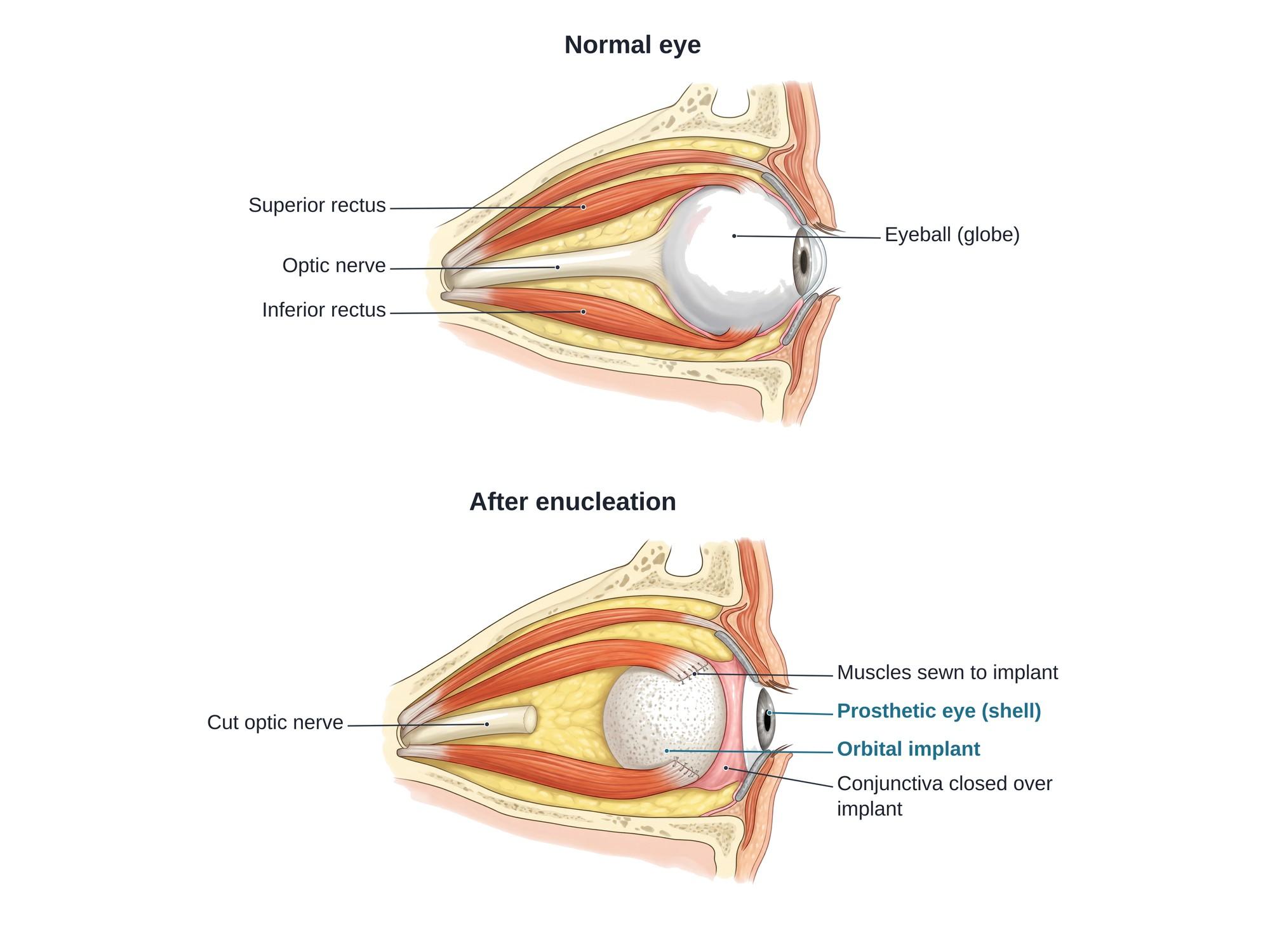
Enucleation is surgery to remove the whole eyeball. The surgeon detaches the eye muscles, cuts the optic nerve and lifts the eye out of the socket. An orbital implant, a round ball, is placed in the eye socket to replace the lost volume. Weeks later, a custom prosthetic eye is fitted behind the eyelids. The illustration above shows a normal eye in the orbit beside the orbit after enucleation.
Why it is done
Enucleation is done when an eye cannot be saved, or when keeping it puts the person at risk. Common reasons are:
- Eye cancer, such as uveal melanoma in adults or retinoblastoma in children, when the tumor is too large for other treatment
- Severe trauma, such as a ruptured globe from a car crash, assault, gunshot or blast, when the eye cannot be repaired
- A blind, painful eye, for example after end-stage glaucoma or a failed retinal detachment repair
- Prevention of sympathetic ophthalmia, a rare immune attack on the healthy eye after a penetrating injury to the other eye
- Severe infection inside the eye that does not respond to treatment
Severe blunt trauma can also cause bleeding inside the eye, such as a hyphema. Most of these eyes heal. Enucleation is a last resort when the eye is destroyed or blind and painful.
How the surgery is done
The operation usually takes 1 to 2 hours under general anesthesia.
- Open the conjunctiva. The surgeon cuts the conjunctiva, the thin clear skin over the white of the eye, in a ring around the cornea.
- Free the eye muscles. The four rectus muscles are tagged with stitches and cut from the eyeball. The two oblique muscles are also released.
- Cut the optic nerve. The surgeon cuts the optic nerve behind the eye and lifts the eyeball out. In cancer cases, a long piece of nerve is taken to check for tumor spread.
- Control bleeding. Pressure and cautery stop bleeding in the socket.
- Place the orbital implant. A round implant is set deep in the socket, inside Tenon capsule, a tough layer of tissue that wrapped the eye.
- Reattach the muscles. The rectus muscles are sewn to the front of the implant, or to a wrap around it, near where they sat on the eye.
- Close in layers. Tenon capsule and then the conjunctiva are closed over the front of the implant.
- Place a conformer. A clear plastic shell is placed behind the eyelids to hold the space for the future prosthetic eye. A pressure patch is put on for a few days.
Hardware
| Part | What it is | Purpose |
|---|---|---|
| Orbital implant | A ball, usually 18 to 22 mm, of porous polyethylene, hydroxyapatite, silicone or acrylic | Replaces the volume of the eye so the socket does not sink |
| Implant wrap | Donor sclera or mesh around some implants | Gives the muscles something to be sewn to |
| Conformer | A clear plastic shell | Holds the shape of the socket while it heals |
| Prosthetic eye | A thin, custom-painted acrylic shell | Matches the look of the other eye and sits behind the lids |
Porous implants let blood vessels and tissue grow into them. This helps hold them in place.
Recovery
- First week: swelling, bruising and aching around the eye. A pressure patch is worn for a few days.
- First weeks: antibiotic and steroid drops or ointment. Most people return to desk work in about 1 to 2 weeks.
- 6 to 8 weeks: an ocularist, a specialist in making artificial eyes, takes a mold of the socket and fits a custom prosthetic eye.
- Long term: the prosthetic eye is removed and cleaned now and then, and polished about once a year. It is usually replaced every few years.
People with one eye can usually drive and work. They need time to adjust to the loss of depth perception and the blind side. Safety glasses are advised to protect the remaining eye.
Risks
- Bleeding and infection in the socket
- Implant exposure, when the conjunctiva wears thin over the implant
- Implant extrusion, when the implant pushes out and must be replaced
- A sunken upper eyelid or a socket that looks hollow over time
- Drooping eyelid (ptosis) or a lower lid that sags
- Poor fit or movement of the prosthetic eye
- Discharge and irritation from the prosthetic eye
- Grief and body image concerns after losing an eye
How enucleation is shown in a personal injury case
Loss of an eye is one of the most serious injuries in a personal injury case. It can follow a car crash, an assault, a workplace accident, a fireworks or BB gun injury, or a delayed diagnosis of eye cancer or infection. A jury may never see the empty socket, because the prosthetic eye can look natural. A clear medical illustration of the normal orbit beside the orbit after enucleation shows what was removed, what replaced it, and why the person will need lifelong prosthetic care. It also helps explain the future costs in a life care plan, such as new prosthetic eyes, socket surgery and protective eyewear. For how these images are used at trial, see demonstrative evidence and medical legal illustration.
FAQ
What does enucleation mean?+
In eye surgery, enucleation means removing the whole eyeball. The surgeon detaches the eye muscles, cuts the optic nerve and lifts the eye out, leaving the eyelids, muscles and the rest of the eye socket in place. The word is also used for removing a whole tumor or cyst in one piece in other parts of the body.
Can you still see after enucleation?+
No, not with that eye. Enucleation removes the eye and cuts the optic nerve, so all vision on that side is lost for good. Most people still see well with the other eye, but they lose depth perception at first and must adjust to a narrower field of view on the side of the removed eye.
How painful is eye enucleation?+
The surgery is done under general anesthesia, so it is not felt. Afterward there is aching, swelling and bruising for a few days to a couple of weeks, usually controlled with pain medicine and cold packs. Many people who had a blind, painful eye feel less pain once it is removed.
What is the difference between enucleation and evisceration?+
Enucleation removes the whole eyeball, including the white sclera, and cuts the optic nerve. Evisceration removes only the contents of the eye and leaves the sclera and muscle attachments in place. Enucleation is chosen when there may be a tumor inside the eye, or when the whole eye must be removed after severe trauma.
What is an orbital implant?+
An orbital implant is a ball placed in the eye socket after the eye is removed. It is often made of porous polyethylene, hydroxyapatite or silicone. It replaces the lost volume, so the socket does not look sunken, and the eye muscles are sewn to it so the prosthetic eye can move.
How long after enucleation can you get a prosthetic eye?+
A clear plastic shell called a conformer is placed at the end of surgery to hold the shape of the socket. The custom prosthetic eye is usually fitted by an ocularist about 6 to 8 weeks later, once the swelling has gone down.
Does a prosthetic eye move?+
Yes, partly. Because the eye muscles are attached to the implant, the implant moves when the other eye moves, and it carries the prosthetic shell with it. The movement is usually less than a natural eye, especially when looking far to the side.
Why would someone need their eye removed?+
The most common reasons are a cancer inside the eye (such as uveal melanoma in adults or retinoblastoma in children), severe trauma that cannot be repaired, a blind and painful eye, and, rarely, a severe infection. After a serious eye injury, removal may also be done to prevent sympathetic ophthalmia, an immune attack on the healthy eye.
Need a surgical illustration for consent or a case?
Describe the operation and get a clear, labeled medical illustration for patient consent, teaching or a case exhibit.
Try Natomy →