Canthoplasty: Lateral Canthoplasty Surgery & Recovery
Canthoplasty tightens the outer corner of the eye by reattaching the lateral canthal tendon to the orbital rim. See the surgery illustrated.
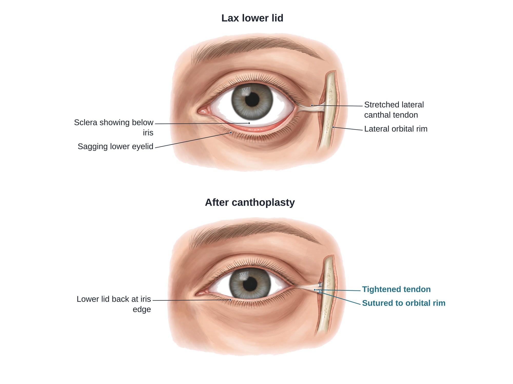
Canthoplasty is eyelid surgery that reshapes and tightens the corner of the eye. Most often it is done at the outer corner, called a lateral canthoplasty. The surgeon releases the lateral canthal tendon, the band that anchors the eyelids to the bone at the outer corner. The lower lid is then shortened and stitched back to the inner side of the bony rim of the eye socket. This pulls a loose, sagging lower lid back against the eye. Canthoplasty is used to treat ectropion, a lid turned outward, and other eyelid problems from aging, nerve palsy, injury or past surgery. It is also done for cosmetic reasons. The illustration above shows a lax lower lid before and after a lateral canthoplasty.
Why it is done
The lower lid rests against the eye like a hammock. It is held at each end by a canthal tendon. When the lateral canthal tendon stretches or tears, the lid sags and pulls away from the eye. Canthoplasty fixes that support. Common reasons are:
- Ectropion, a lower lid that sags and turns outward, leaving the eye dry, red and watery
- Entropion, a lid that turns inward so the lashes rub the eye. The repair often includes tightening the outer corner.
- Lower eyelid laxity, a loose lid that does not stay against the eye
- Lower eyelid retraction, a lid that is pulled down, often after earlier lower lid surgery (blepharoplasty)
- Facial nerve palsy, where the muscle that holds the lid up no longer works
- Injury to the outer corner, such as a cut, a dog bite or a broken cheekbone that tears the tendon
- Support during other surgery, such as lower lid blepharoplasty or eye socket fracture repair, to prevent the lid from pulling down
- Cosmetic reshaping, sometimes called an eye corner lift or almond eye surgery
How the surgery is done
The most common type of lateral canthoplasty is the lateral tarsal strip procedure. It usually takes about 30 to 60 minutes per side, as outpatient surgery under local anesthesia with sedation.
- Numbing. The surgeon injects local anesthetic around the outer corner of the eye.
- Lateral canthotomy. A short horizontal cut is made through the skin and tissue at the outer corner of the eye.
- Cantholysis. The lower limb of the lateral canthal tendon is cut free from the bone. The lower lid is now loose and can be pulled outward.
- Measuring. The surgeon pulls the lid toward the bone and marks how much extra length must be removed.
- Making the tarsal strip. At the outer end of the lid, the skin, the lash edge and the inner lining are trimmed away. This leaves a short, firm strip of the tarsal plate, the stiff tissue that gives the lid its shape. Any extra length is removed.
- Reattaching to bone. The strip is stitched to the periosteum, the tough lining of the bone, on the inner side of the lateral orbital rim. This spot is a small bump inside the rim called the lateral orbital tubercle. Fixing the lid inside the rim keeps it pressed against the eye.
- Rebuilding the corner. The upper and lower lids are joined again to form a sharp outer corner.
- Closing. The muscle and skin are closed with fine stitches.
In a canthopexy, a simpler option, the tendon is not cut. The surgeon passes a stitch through the existing tendon and fixes it to the inside of the rim to tighten it.
Recovery
- Swelling and bruising around the eye for 1 to 2 weeks
- Cold packs for the first 2 to 3 days
- Antibiotic ointment on the incision for about a week
- Sleeping with the head raised for the first few days
- No heavy lifting, bending or swimming for about 2 weeks
- Stitches removed or dissolved within about a week
The outer corner often looks tight and slightly high at first. It relaxes over the following weeks. The final position settles over about 2 to 3 months.
Risks
- Bleeding and bruising. Rarely, bleeding behind the eye puts pressure on the optic nerve. This is an emergency.
- Infection
- Asymmetry between the two eyes
- A rounded or webbed outer corner instead of a sharp angle
- A lid that is too tight, which can slide under a prominent eye instead of resting against it
- A lid that is still too loose, or loosens again over time
- Stitch problems, such as a stitch poking through or a small lump of scar tissue around it
- Dry eye or irritation while the lid heals
- Visible scar at the outer corner, usually faint once healed
How canthoplasty is shown in a personal injury case
Canthoplasty comes up in injury cases in two ways. First, it can be the repair after trauma. A car crash, a fall, an assault or a dog bite can tear the lateral canthal tendon or break the cheekbone, leaving a sagging lid and a rounded, misshapen outer corner. The injured person may need one or more canthoplasties, and the change in the shape of the eye can be permanent. Second, it can be a revision after earlier eyelid surgery. A lower lid blepharoplasty that removes too much skin or weakens the lid can pull it down, and canthoplasty is part of the fix. A clear medical illustration of the lax lid before surgery and the tightened lid after shows a jury what the tendon does, why the lid sagged, and what the surgeon had to rebuild. For how these images are used at trial, see demonstrative evidence and medical legal illustration.
FAQ
What is lateral canthoplasty?+
Lateral canthoplasty is eyelid surgery at the outer corner of the eye. The surgeon releases the lateral canthal tendon, the band that anchors the eyelids to the bone, then shortens the lower lid and stitches it back to the inside of the orbital rim. It tightens a loose lower lid and can reshape the corner of the eye.
What is the difference between a canthopexy and a canthoplasty?+
In a canthopexy, the surgeon tightens the existing tendon with stitches and does not cut it. In a canthoplasty, the tendon is cut and released, the lid is shortened, and the corner is rebuilt and reattached to the bone. A canthopexy suits mild looseness. A canthoplasty gives a stronger, longer lasting fix for a very loose or out of place lid.
Is canthoplasty permanent?+
The repair is meant to last. The new attachment heals as scar to the bone, and most results hold for many years. Aging can loosen the tissues again over time, and some people need a second operation, especially when the lid was very loose or scarred.
What is an eye corner lift?+
Eye corner lift is a cosmetic name for lateral canthoplasty or canthopexy done to change the shape of the eye. Raising or lengthening the outer corner gives a more almond or upswept shape. It is the same basic operation as the medical repair, with the same risks.
How long is canthoplasty recovery?+
Most people have swelling and bruising for 1 to 2 weeks and return to desk work in about a week. Skin stitches are removed or dissolve in about a week. The corner of the eye can look tight or slightly high at first. The final shape settles over about 2 to 3 months.
Does canthoplasty hurt?+
The surgery is usually done under local anesthesia with sedation, so it should not hurt during the operation. Afterward, most people have soreness and tightness at the outer corner for several days. It is usually controlled with cold packs and simple pain relievers.
How much does canthoplasty cost?+
Cost depends on the surgeon, the region, the facility and whether the surgery is done for a medical problem or for looks. When it treats a medical problem such as ectropion or eyelid retraction, insurance may cover it. Cosmetic canthoplasty is usually paid out of pocket.
What is a lateral canthotomy?+
A lateral canthotomy is a small cut through the outer corner of the eye. It is the first step of a canthoplasty. Done with a cantholysis, which releases the lower tendon, it is also an emergency procedure to relieve dangerous pressure behind the eye after bleeding in the orbit.
Need a surgical illustration for consent or a case?
Describe the operation and get a clear, labeled medical illustration for patient consent, teaching or a case exhibit.
Try Natomy →