Ectropion: Causes, Types & Eyelid Repair Surgery
Ectropion is a lower eyelid that sags and turns outward, leaving the eye dry, red and watery. See it illustrated beside a normal eyelid.
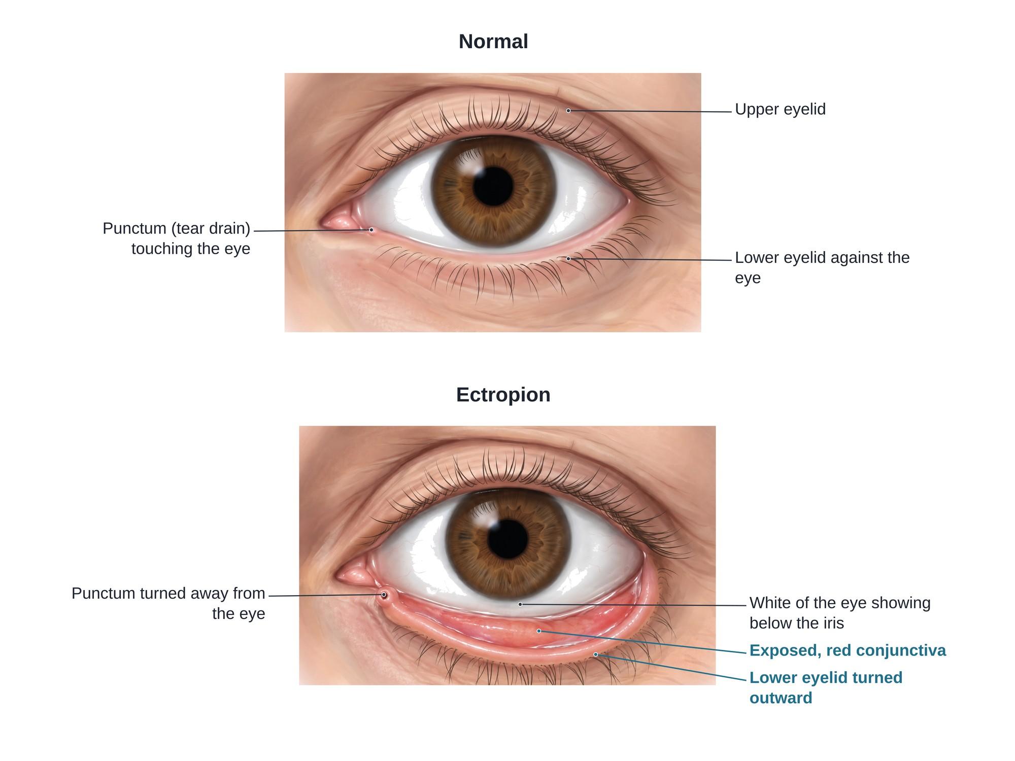
Ectropion is a condition in which the eyelid, usually the lower lid, sags and turns outward, away from the eye. The pink inner lining of the lid is exposed, and the eye cannot close or stay moist properly. The result is a dry, red, irritated eye that often waters. Ectropion is most common in older adults. It can also follow facial nerve palsy, burns or scarring from injury or surgery. The illustration above shows a normal lower eyelid beside an eyelid with ectropion.
The anatomy
The lower eyelid is a thin flap held against the eyeball like a hammock. Its layers, from front to back, are:
- Skin, the thinnest skin in the body
- Orbicularis oculi muscle, which closes the eye
- Tarsal plate, a firm strip of tissue that gives the lid its shape
- Conjunctiva, a moist pink lining that faces the eye
The lid is anchored at both ends by the medial canthal tendon near the nose and the lateral canthal tendon at the outer corner. These keep the lid tight against the eye.
Near the inner corner, each lid has a tiny opening called the punctum. It sits against the eye and drains tears into the tear duct and nose. When the lid turns out, the punctum lifts away from the eye, and tears spill onto the cheek.
Types
| Type | Cause | Notes |
|---|---|---|
| Involutional (age-related) | Stretched canthal tendons and weak muscle | The most common type |
| Paralytic | Facial nerve (cranial nerve VII) palsy, such as Bell palsy, stroke, tumor or nerve injury | The orbicularis muscle can no longer hold the lid up |
| Cicatricial (scarring) | Scarring or shortage of skin from burns, cuts, sun damage, skin disease or past eyelid surgery | The skin pulls the lid down and out |
| Mechanical | A growth, swelling or heavy cheek pulling the lid down | Often improves when the cause is removed |
| Congenital | Present from birth, often with other eyelid problems | Rare |
Causes
- Aging, which loosens the tendons and thins the muscle
- Facial nerve palsy from Bell palsy, stroke, skull base surgery or a facial injury
- Burns to the face, including chemical and thermal burns
- Eyelid and cheek lacerations, including dog bites and car crash injuries
- Facial fractures, such as a broken cheekbone or eye socket floor, and the scarring after their repair
- Past eyelid surgery, such as lower lid blepharoplasty where too much skin was removed
- Skin cancer on the lid or its removal
- Long-term sun damage and skin conditions that tighten the skin
Symptoms
- Watery eye (epiphora), with tears running onto the cheek
- Dry, gritty or burning feeling
- Redness of the eye and of the exposed lid lining
- Light sensitivity
- Mucus discharge and crusting
- A visible gap between the lid and the eye, and a red rim of tissue along the lower lid
- Blurred vision if the cornea becomes dry or damaged
Diagnosis
An eye doctor or oculoplastic surgeon diagnoses ectropion on a routine exam. Common tests are:
- Snap-back test. The doctor pulls the lid down and lets go. A healthy lid snaps back at once. A loose lid returns slowly.
- Lid distraction test. The doctor pulls the lid away from the eye. More than about 6 mm suggests a loose lid.
- Checking the canthal tendons at both corners for looseness
- Looking for scarring by asking the patient to look up and open the mouth, which pulls a scarred lid down further
- Testing the facial nerve for weakness
- Staining the cornea with fluorescein dye to look for dry spots or scratches
Treatment
Non-surgical care protects the eye while waiting for surgery or when surgery is not possible:
- Lubricating eye drops by day and ointment at night
- Taping the lower lid up at night
- Treating any infection or inflammation
Ectropion repair surgery is the lasting fix. The surgeon picks the operation that matches the cause:
- Lateral tarsal strip. The surgeon shortens the lower lid at the outer corner and stitches it to the bone of the eye socket rim. This is the most common repair for age-related ectropion. Tightening the outer corner this way is a form of canthoplasty, done through a small cut at the outer corner.
- Medial spindle procedure. A small diamond of the inner lid lining is removed to turn the inner lid and punctum back toward the eye.
- Medial canthal tendon repair when the inner end of the lid is loose.
- Skin graft or local flap for scarring, to add the skin that is missing.
- Gold weight or other procedures for facial nerve palsy, to help the eye close.
Most repairs are done as outpatient surgery under local anesthesia with light sedation. They take about 30 to 60 minutes per lid.
Recovery
- Bruising and swelling for 1 to 2 weeks
- Antibiotic ointment for about a week
- Cold packs in the first few days
- No heavy lifting or swimming for about 1 to 2 weeks
- Stitches removed or dissolved within about a week
Most people notice less tearing and irritation within weeks. The final lid position settles over a few months.
Complications
Of the condition, if not treated:
- Exposure keratopathy, dry and damaged cornea
- Corneal ulcers and infection, which can scar the cornea and harm vision
- Thickened, hardened lid lining from long exposure
- Skin irritation of the cheek from constant tears
Of surgery:
- Bleeding, infection and scarring
- A lid that is too tight or too loose
- A change in the shape of the outer corner of the eye
- Return of the ectropion over time
How ectropion is shown in a personal injury case
Ectropion is often the long-term result of an injury. A facial burn, a deep eyelid cut, a dog bite or a broken cheekbone can scar the lower lid and pull it outward. A facial nerve injury can leave the lid too weak to stay up. Ectropion is also a known complication of lower eyelid cosmetic surgery, where the question is whether too much skin was removed. A clear medical illustration of a normal eyelid beside an eyelid with ectropion shows a jury why the injured person has a red, watery, painful eye, why it puts their vision at risk, and why they may need more surgery. For how these images are used at trial, see demonstrative evidence and medical legal illustration.
FAQ
What is the cause of ectropion?+
The most common cause is aging. The tendons and muscle that hold the lower lid against the eye stretch and weaken over time. Other causes are facial nerve palsy (such as Bell palsy or a nerve injury), scarring of the eyelid skin from burns, cuts or surgery, growths that pull the lid down, and, rarely, a birth defect.
How do you treat ectropion?+
Mild ectropion may be managed with lubricating eye drops and ointment, and taping the lid at night. Most cases need surgery to fix it for good. The most common operation tightens the lower lid at the outer corner (a lateral tarsal strip). Scarring may also need a skin graft, and nerve palsy may need other procedures.
What happens if ectropion is left untreated?+
The exposed eye can stay dry, red and irritated. Over time, the cornea can get scratches, ulcers or scarring, which can harm vision. The inner lining of the lid can thicken and harden, and constant tearing can irritate the skin of the cheek.
What is the difference between ectropion and entropion?+
In ectropion, the eyelid turns outward, away from the eye. In entropion, the eyelid turns inward, so the lashes rub against the eye. Both are most common in the lower lid of older adults, and both are usually fixed with eyelid surgery.
Is eyelid ectropion the same as cervical ectropion?+
No. They share a name because both mean tissue turned outward. Cervical ectropion is a harmless change on the cervix in the female reproductive tract. This page is about ectropion of the eyelid.
Why does ectropion cause watery eyes?+
Tears drain through a tiny opening called the punctum near the inner corner of each eyelid. When the lower lid turns out, the punctum no longer touches the eye, so tears spill over the lid onto the cheek. At the same time, the exposed eye gets dry and makes more reflex tears.
How long is recovery from ectropion surgery?+
Most people have bruising and swelling for 1 to 2 weeks. Stitches are often removed or dissolve within about a week. Most return to normal activities in 1 to 2 weeks, and the final lid position settles over a few months.
Can ectropion come back after surgery?+
Yes, sometimes. The lid can loosen again with age, or scarring can pull it down again. Most repairs last for years, but some people need a second operation.
Need this illustration for a patient or a case?
Describe the injury or upload a scan and get a clear, labeled medical illustration for a consult, a chart or a case exhibit.
Try Natomy →