Nutcracker Syndrome: Causes, Symptoms & Treatment
Nutcracker syndrome is compression of the left renal vein between the aorta and the superior mesenteric artery. See it illustrated in a front view.
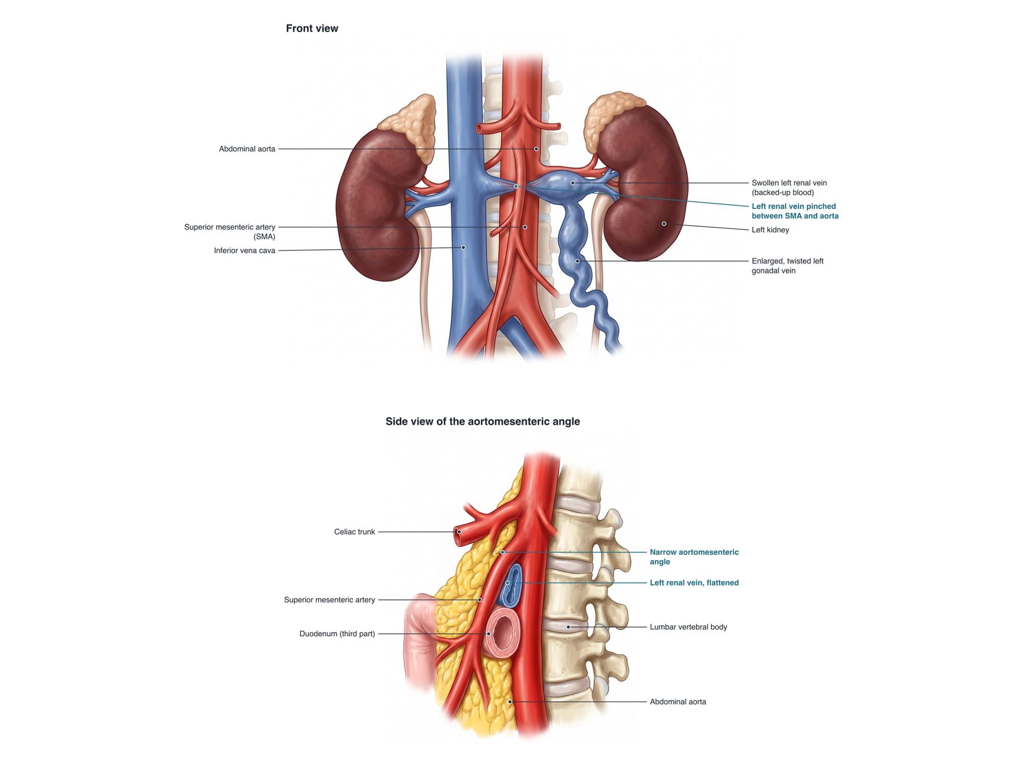
Nutcracker syndrome is compression of the left renal vein as it passes between the abdominal aorta behind it and the superior mesenteric artery (SMA) in front of it. Like a nut in a nutcracker, the vein is squeezed in the angle between the two arteries. Blood backs up toward the left kidney, raising the pressure in the vein. This can cause blood in the urine, left flank pain and enlarged pelvic veins. When the narrowing is seen on a scan without symptoms, it is called the nutcracker phenomenon. The illustration above shows the left renal vein squeezed between the SMA and the aorta, with the swollen vein and enlarged gonadal vein behind the pinch.
The anatomy
The left kidney sits farther from the inferior vena cava than the right one. So the left renal vein is long. It crosses the midline to reach the vena cava, passing in front of the aorta and just below the origin of the SMA. The SMA normally leaves the front of the aorta at an angle of about 40 to 60 degrees. A pad of fat and lymph tissue fills the space and protects the vein. The left gonadal vein (ovarian or testicular vein) and left adrenal vein drain into the left renal vein. So pressure in the renal vein spreads into them. For more on these organs, see abdominal organs anatomy.
Types
| Type | What happens |
|---|---|
| Anterior nutcracker | The left renal vein is squeezed between the SMA and the aorta. This is the classic and most common form. |
| Posterior nutcracker | A left renal vein that runs behind the aorta is squeezed between the aorta and the spine. |
| Combined | A duplicated vein, with one branch in front of the aorta and one behind, compressed in both places. |
Causes
- A narrow SMA angle: the SMA leaves the aorta at a sharp angle, often under 25 to 35 degrees.
- Low body fat or weight loss: less fat between the arteries, common in thin teenagers and young adults.
- Rapid growth in height during adolescence, which stretches the vessels.
- Increased lumbar lordosis (a deep curve of the lower back), which pushes the aorta forward.
- Less common: a tumor, enlarged lymph nodes, an aortic aneurysm, or a kidney that drops when standing.
- Pregnancy can bring out symptoms.
Symptoms
- Blood in the urine (hematuria), often the first sign, visible or only on testing
- Left flank or abdominal pain, often worse with standing, sitting or exercise
- Protein in the urine, especially after standing (orthostatic proteinuria)
- Pelvic pain and pelvic varicose veins in women, from reflux down the left ovarian vein (pelvic congestion)
- Left-sided varicocele in men, an enlarged vein network in the scrotum
- Pain during or after intercourse, painful periods, and fatigue in some patients
- Some have no symptoms at all
Diagnosis
Diagnosis is often delayed because the symptoms overlap with many other conditions. Urine tests look for blood and protein. Cystoscopy, a camera look inside the bladder, can show blood coming from the left ureter. Doppler ultrasound measures how fast blood moves through the narrowed vein compared with the wider part near the kidney. A large speed difference supports the diagnosis. CT or MR venography shows the narrowed vein, the angle of the SMA, and any enlarged gonadal or pelvic veins. A beak-shaped narrowing of the vein is a common finding. Venography with pressure measurements is the most direct test. A catheter measures the pressure on each side of the narrowing. A pressure difference of more than about 3 mmHg supports nutcracker syndrome. Other causes of blood in the urine, such as stones and tumors, must also be ruled out.
Treatment
- Watchful waiting: for mild symptoms, especially under 18, since many improve as the body matures and fat builds around the vessels
- Weight gain in very thin patients, guided by a doctor or dietitian
- Medicines: ACE inhibitors for protein in the urine, and pain medicine
- Left renal vein transposition: the most common open surgery. The vein is cut and reattached lower on the vena cava, away from the SMA.
- Gonadal vein transposition or bypass: the gonadal vein is used to drain the kidney around the blockage
- Kidney autotransplantation: the kidney is moved lower in the pelvis with new vessel connections
- Endovascular stent: a metal tube placed in the vein. It is less invasive, but stents can move, so it is used selectively.
- Gonadal or pelvic vein embolization: closing enlarged pelvic veins to treat pelvic congestion
Recovery
After open vein transposition, most people stay in hospital for a few days and return to light activity in about 4 to 6 weeks. Blood in the urine often stops within weeks. Pain can take several months to settle, and some people keep some pain. After a stent, recovery is faster, often a week or two, but follow-up scans check its position.
Complications
- Anemia from ongoing blood loss in the urine
- Chronic left flank and pelvic pain
- Pelvic congestion syndrome and varicose veins of the pelvis, vulva or legs
- Left varicocele and, in some men, reduced fertility
- Blood clots in the left renal vein (rare)
- After treatment: stent movement or blockage, and narrowing at the new vein connection
How nutcracker syndrome is shown in a personal injury case
Nutcracker syndrome comes up most often in medical malpractice claims about a delayed diagnosis. Patients may see several doctors for blood in the urine, flank pain or pelvic pain before anyone checks the left renal vein. Complications of treatment, such as a stent that moves to the heart, can also lead to claims. It is not usually caused by trauma, but rapid weight loss after an injury or illness can bring it out. A front view illustration like the one above lets a jury see why the vein is squeezed and how the blood backs up. For how these images are used at trial, see demonstrative evidence and medical legal illustration.
FAQ
What are the urinary symptoms of nutcracker syndrome?+
The main one is blood in the urine (hematuria). It can be visible, making the urine pink, red or cola-colored, or found only on a urine test. Some people also have protein in the urine (proteinuria), which may be worse after standing for a long time.
Can nutcracker syndrome kill you?+
It is very rarely life-threatening. Most people with mild symptoms do well, and many children outgrow it. Severe cases can cause heavy bleeding into the urine, anemia and chronic pain, which need treatment. Any heavy or persistent bleeding should be checked promptly.
How rare is nutcracker syndrome?+
The true rate is not known. Mild narrowing of the left renal vein is common on scans and often causes no symptoms. That finding is called the nutcracker phenomenon. Nutcracker syndrome, with symptoms, is much less common. It is most often found in thin young adults and teenagers, and more often in women.
What are vascular compression syndromes?+
They are conditions where a blood vessel or nearby structure is squeezed by another. Examples include nutcracker syndrome (left renal vein), May-Thurner syndrome (left iliac vein), superior mesenteric artery syndrome (duodenum) and median arcuate ligament syndrome (celiac artery). Some people have more than one.
Can nutcracker syndrome cause pain after eating?+
Nutcracker syndrome mainly causes left flank and pelvic pain. Pain after eating, with nausea and early fullness, points more to superior mesenteric artery syndrome. That condition involves the same narrow angle and sometimes occurs together with nutcracker syndrome.
Does diet help nutcracker syndrome?+
In thin patients, gaining a healthy amount of weight can add fat around the SMA. That can widen the angle and ease the compression, especially in teenagers. Diet changes should be guided by a doctor or dietitian.
Can nutcracker syndrome cause headaches?+
Headaches are not a typical feature. Some patients with several compression syndromes report a wide range of symptoms, including fatigue and headaches, but a direct link has not been proven. Other causes of headache should be checked.
How is nutcracker syndrome treated?+
Mild cases, especially in young people, are often watched for 1 to 2 years because many improve. For severe or lasting symptoms, options include moving the left renal vein lower on the vena cava (transposition), a gonadal vein bypass, kidney autotransplantation, or sometimes a stent.
Need this illustration for a patient or a case?
Describe the injury or upload a scan and get a clear, labeled medical illustration for a consult, a chart or a case exhibit.
Try Natomy →