Megalocornea: Causes, Signs & Diagnosis vs Normal Eye
Megalocornea is a cornea over 13 mm wide, present from birth, with a deep front chamber and normal eye pressure. See it illustrated beside a normal eye.
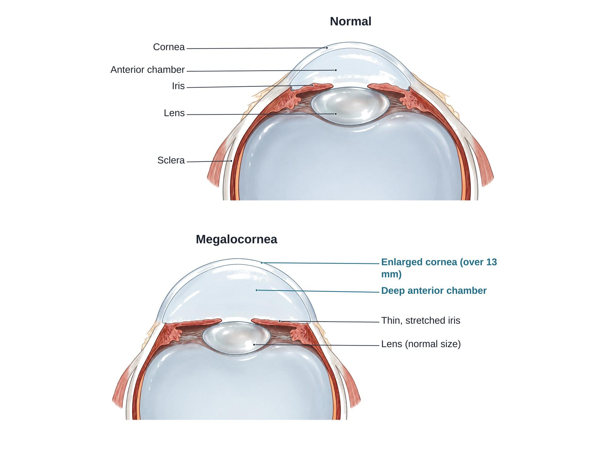
Megalocornea is a cornea that is abnormally wide, 13 mm or more across, while the cornea stays clear and of normal thickness. It is present from birth, usually affects both eyes, and does not get bigger after early childhood. The front chamber of the eye is deeper than normal, but the eye pressure is normal. Most cases are inherited and occur in boys. The illustration above shows a normal eye beside an eye with megalocornea.
The anatomy
The cornea is the clear dome at the front of the eye. It does about two thirds of the eye's focusing. In an adult, it is about 11 to 12 mm wide side to side. Its edge meets the white sclera at a band called the limbus.
Behind the cornea is the anterior chamber, a space filled with clear fluid called aqueous. At the back of this chamber are the colored iris and the opening in its center, the pupil. Behind the iris sits the lens, held in place by fine fibers called zonules that attach to the ciliary body.
In megalocornea, the whole ring at the front of the eye is too wide. The cornea is larger, the anterior chamber is deeper, and the iris and ciliary body are stretched. The eye behind the lens is usually a normal size.
Types
| Type | What it is | Notes |
|---|---|---|
| X-linked (isolated) megalocornea | Large cornea alone, from a CHRDL1 gene change | The most common type. About 90 percent are male. |
| Autosomal megalocornea | Large cornea inherited from one or both parents | Rare. Affects both sexes. |
| Anterior megalophthalmos | Large cornea plus a stretched iris, ciliary body and loose lens | Higher risk of cataract and lens dislocation |
| Syndromic megalocornea | Part of a body-wide condition | Seen with Marfan syndrome, Down syndrome, Alport syndrome and others |
Megalocornea vs congenital glaucoma
The main concern in a baby with large corneas is congenital glaucoma, sometimes called buphthalmos ("ox eye"). It needs urgent surgery.
| Feature | Megalocornea | Congenital glaucoma |
|---|---|---|
| Cornea | Clear, normal thickness | Often cloudy and swollen, with breaks in its back layer (Haab striae) |
| Eye pressure | Normal | High |
| Eye length | Normal | Longer than normal |
| Optic nerve | Healthy | Cupped, damaged |
| Symptoms | Usually none | Tearing, light sensitivity, squeezing the eyes shut |
| Progression | Stable | Gets worse without treatment |
Causes
- X-linked inheritance. Changes in the CHRDL1 gene are the most common cause. Mothers who carry the gene usually have normal eyes. Their sons have a 1 in 2 chance of being affected.
- Other genes, rarely, passed down in families in other patterns.
- Syndromes, such as Marfan syndrome, Down syndrome, Alport syndrome, Ehlers-Danlos syndrome and megalocornea with intellectual disability (Neuhauser syndrome).
Symptoms
Many people have no symptoms. Others notice:
- Eyes that look big or "bright" in a baby or child
- Blurry vision from nearsightedness or astigmatism
- Glare, from a thin iris that lets light through
- A wobbly iris when the eye moves (iridodonesis)
Diagnosis
An eye doctor measures the width of the cornea with a ruler or caliper, or with a scan. The doctor also checks:
- Eye pressure, which is normal in megalocornea
- Corneal thickness, which is normal (it is thin in keratoglobus, another cause of a large-looking cornea)
- Anterior chamber depth, which is deeper than normal
- Eye length, measured with ultrasound biometry, which is normal
- The optic nerve, which is healthy
- The lens, for early cataract or a loose lens
In a baby, the exam may be done under anesthesia. Genetic testing and a check for related syndromes may follow.
Treatment
Megalocornea itself does not need treatment. Care focuses on vision and on problems that can come later.
- Glasses or contact lenses for nearsightedness and astigmatism
- Amblyopia treatment, such as patching, if one eye sees much worse in childhood
- Cataract surgery when a cataract forms. These eyes have a deep chamber, weak zonules and a large capsule, so surgery needs special lenses and planning.
- Glaucoma treatment if eye pressure rises later in life
Recovery
There is no recovery period, because megalocornea is a lifelong shape of the eye, not an injury. Most people have useful vision. Yearly eye exams are advised to watch for cataract, lens dislocation and glaucoma.
Complications
- Early cataract, often in the 30s to 50s
- Lens subluxation or dislocation, when weak zonules let the lens slip out of place
- Pigment dispersion and glaucoma, when the stretched iris sheds pigment that blocks the eye's drain
- Retinal detachment, in some patients
- Hard cataract surgery, with a higher risk of complications
How megalocornea is shown in a personal injury case
Megalocornea is not caused by trauma. It comes up in medical malpractice cases about a missed diagnosis in a baby. Large, cloudy corneas in an infant may be congenital glaucoma, which needs prompt surgery to save sight. If a doctor assumes the eyes are just large, or misses the signs of glaucoma, the child can lose vision. It can also come up after cataract surgery in an adult, where the deep chamber and weak lens support make complications more likely and the question is whether the surgeon planned for them. A clear medical illustration of a normal eye beside an eye with megalocornea shows a jury where the cornea, chamber and lens sit, and why this eye is different. For how these images are used at trial, see demonstrative evidence and medical legal illustration.
FAQ
Why does megalocornea occur?+
Most cases are inherited. The most common form is X-linked, caused by changes in the CHRDL1 gene, so it mainly affects boys and is passed through the mother. Researchers think the ring of tissue that forms the cornea grows too wide early in development. Less often it is part of a syndrome, such as Marfan syndrome or Down syndrome.
What size cornea is megalocornea?+
A normal adult cornea is about 11 to 12 mm wide side to side. Megalocornea means a horizontal diameter of 13 mm or more after about age 2, or 12.5 mm or more in a newborn. The cornea stays clear and of normal thickness.
Is megalocornea the same as congenital glaucoma?+
No, but they can look alike in a baby. In congenital glaucoma, high eye pressure stretches the whole eye, so the cornea gets cloudy, the eye gets longer and the optic nerve is damaged. In megalocornea, the eye pressure is normal, the cornea is clear and the eye behind the cornea is a normal length. An eye doctor checks all of these to tell them apart.
Does megalocornea affect vision?+
Vision is often good. Many people need glasses for nearsightedness (myopia) or astigmatism, a cornea that is not evenly curved. Problems can come later from early cataract, a loose lens or glaucoma, so regular eye exams matter.
Does megalocornea get worse over time?+
The cornea itself does not keep growing after early childhood. The condition is called non-progressive. But adults with megalocornea have a higher risk of cataract in their 30s to 50s, lens dislocation and some types of glaucoma.
Is megalocornea in one eye or both eyes?+
Isolated megalocornea is almost always in both eyes and fairly even. A large cornea in only one eye is more suspicious for glaucoma or another cause and needs a careful exam.
How is megalocornea treated?+
There is no treatment to shrink the cornea, and none is needed. Care is glasses or contact lenses to correct vision, and lifelong eye exams to catch cataract, lens problems or glaucoma early. Cataract surgery in these eyes is harder and should be planned by an experienced surgeon.
Can megalocornea be seen from the side?+
Yes. From the side, the clear dome of the cornea looks larger and the front chamber of the eye looks deeper than usual. The colored iris may look far back from the front of the eye and may wobble slightly when the eye moves, a sign called iridodonesis.
Need this illustration for a patient or a case?
Describe the injury or upload a scan and get a clear, labeled medical illustration for a consult, a chart or a case exhibit.
Try Natomy →