Basics of Human Anatomy: A Complete Guide for 2026
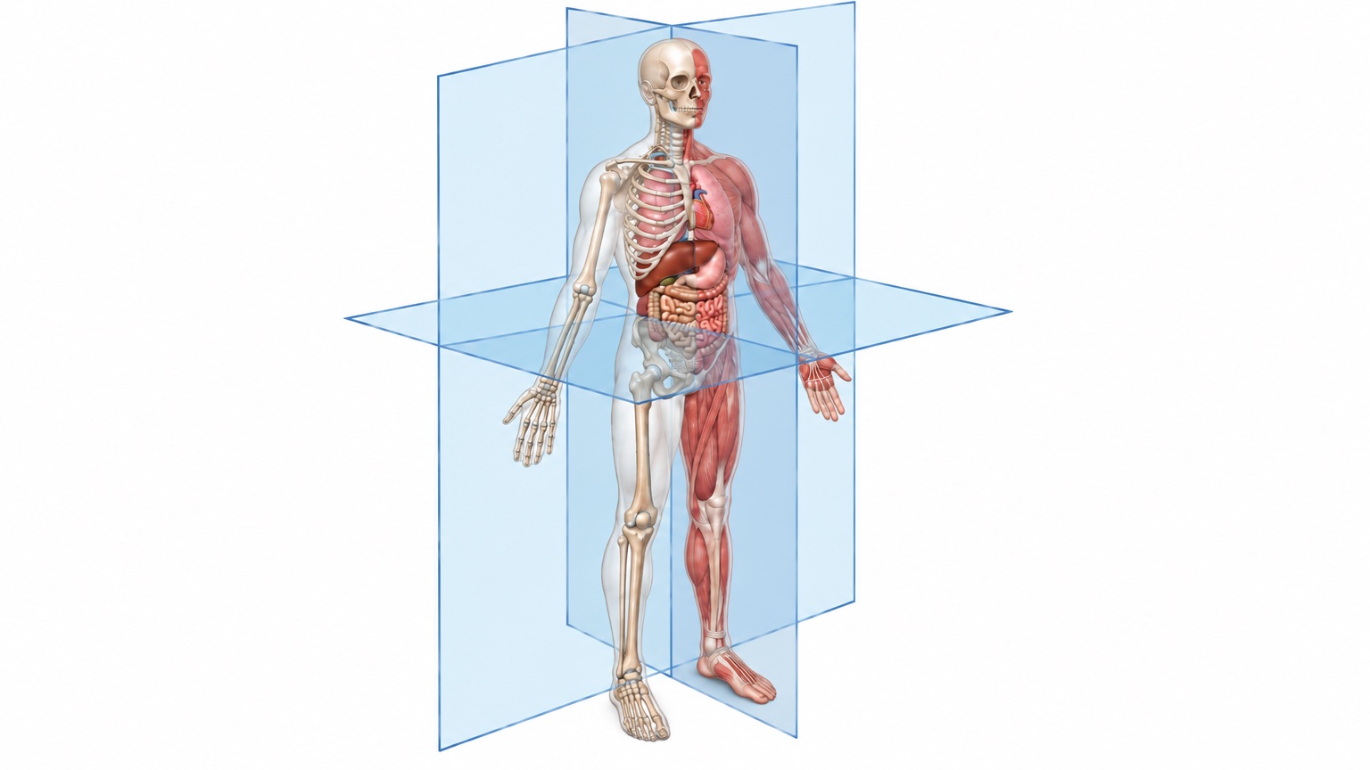 Human body in anatomical position, split to show the skeleton on one side and the muscles on the other, with the heart, lungs, liver, stomach, and intestines visible and the sagittal, coronal, and transverse anatomical planes passing through the body
You're probably here because a static anatomy diagram isn't doing the job.
A surgeon needs to explain a procedure to a patient. A researcher is building slides for a grant presentation. A malpractice attorney wants to show how one structure affected another during an injury. In each case, the problem is the same: anatomy isn't just a list of parts. It's motion, position, force, scale, and consequence.
That's why the basics of human anatomy matter far beyond the classroom. If you only memorize names, you'll miss the part that matters most in practice: how structures relate to one another in space and how those relationships change function. A tendon doesn't matter because it has a name. It matters because it pulls across a joint, redirects force, and can fail in a recognizable pattern.
Traditional medical visuals often flatten those relationships into labels and arrows. Clear anatomy communication requires more than decoration. It requires accuracy, context, and a sense of movement.
Introduction Why Anatomy Is More Than Memorization
Many readers first meet anatomy as a vocabulary burden. You're handed a page of terms, a diagram full of labels, and the unspoken assumption that if you can point to a structure, you understand it. In real clinical and research work, that falls apart quickly.
A physician doesn't ask, “Can I name this muscle?” The practical question is, “If this muscle weakens, what movement changes?” A radiologist doesn't only identify a structure on an image. They interpret whether its position, shape, or relationship to nearby tissues helps explain a symptom. A scientist preparing a figure for publication has the same challenge in another form. The figure must show function, not just form.
The adult human skeleton has 206 bones, 80 in the axial skeleton and 126 in the appendicular skeleton, according to OpenStax Anatomy and Physiology. Babies start with more, often estimated at around 270, because many bones fuse during childhood and adolescence. That fact is useful, but only if you connect it to the living system. Bones aren't museum pieces. They create levers, protect organs, and provide attachment points for muscles.
Practical rule: If an anatomical fact doesn't help you explain movement, pathology, imaging, or procedure, you probably haven't learned it deeply enough yet.
There's a historical reason anatomy can feel split between memorization and understanding. Systematic anatomical study took shape in classical antiquity with physicians such as Herophilus and Galen, Andreas Vesalius rebuilt it on direct human dissection in 1543, and the microscope then opened up a new scale entirely. Robert Hooke coined the term "cell" in 1665, and Antonie van Leeuwenhoek used his single-lens microscopes to describe red blood cells and sperm cells. This historical timeline of anatomy in the Sultan Qaboos University Medical Journal traces how the field developed. Anatomy grew by expanding perspective, not by collecting labels.
That same lesson still applies. The basics of human anatomy become useful when you treat the body as a coordinated, three-dimensional, changing structure.
The Language of Anatomy Positions Planes and Directions
If anatomy had a grammar, this would be it. Before you can interpret a scan, describe a lesion, or explain a surgical route, you need a common spatial language.
Anatomical position as the shared starting point
Anatomists describe the body from a standard reference stance called the anatomical position. In plain language, picture a person standing upright, facing forward, arms at the sides, with the palms facing forward. This isn't how people usually stand. It's used because everyone in medicine needs the same reference point.
If you skip this step, directional language becomes slippery. “Front,” “back,” “inside,” and “outside” can all shift with posture. Anatomical position fixes that problem.
 A diagram illustrating anatomical terminology, including body positions, anatomical planes, and essential directional terms for medical study.
For learners who want extra reinforcement of the vocabulary itself, the anatomical terminology chapter of OpenStax Anatomy and Physiology is a free, peer-reviewed reference that covers anatomical position, directional terms, and body planes with clear figures.
Planes as slices through the body
The three major anatomical planes help you mentally cut the body into sections.
| Plane | What it divides | Everyday way to picture it |
|---|---|---|
| Sagittal | Left and right portions | Slicing an apple from front to back |
| Coronal | Front and back portions | Slicing an apple to separate the front from the rear |
| Transverse | Upper and lower portions | Cutting across the middle into top and bottom |
These planes matter because modern imaging uses them constantly. A CT or MRI image makes more sense when you know whether you're looking at a side division, a front-back division, or a horizontal cross-section.
A common confusion appears here. People often think a plane is a physical structure. It isn't. It's an imaginary reference surface used for description and imaging. Once that clicks, cross-sectional anatomy becomes much easier to interpret.
Directional terms as your compass
Directional terms are the body's compass points. They let clinicians describe location with precision.
- Superior means toward the head.
- Inferior means toward the feet.
- Anterior means toward the front.
- Posterior means toward the back.
- Medial means toward the midline.
- Lateral means away from the midline.
- Proximal means closer to the trunk or to a limb's point of attachment.
- Distal means farther from the trunk or from a limb's point of attachment.
Here's where learners often stumble: medial and lateral are not the same as left and right. A structure can be medial on either side of the body if it lies closer to the center.
When a clinician says pain radiates from the shoulder to the distal forearm, every word is doing spatial work.
Use the terms aloud when you study. Don't just recognize them on a page. Say, “The thumb is lateral to the little finger in anatomical position,” and notice how the sentence forces you to think spatially rather than visually guessing.
Mapping the Body An Overview of Major Organ Systems
When students first learn anatomy, they often meet the body as a pile of disconnected chapters. Skin in one week. Bones in another. Heart later. That's tidy for a syllabus, but it's not how the body works.
Think in systems, not isolated organs
A more useful mental model is a city. Structures have specialized jobs, but the city only functions because those jobs are coordinated. Roads, power, communication, waste removal, and physical buildings don't operate independently. Neither does the body.
Most textbooks organize the human body into 11 organ systems: integumentary, skeletal, muscular, nervous, endocrine, cardiovascular, lymphatic, respiratory, digestive, urinary, and reproductive, as laid out in OpenStax Anatomy and Physiology. Some sources merge the skeletal and muscular systems into a single musculoskeletal system and count 10, but the structures are the same either way. For scale, the adult skeleton has 206 bones, and at rest each ventricle of the heart pumps a mean of about 5.25 liters of blood per minute (normal range 4 to 8 liters), according to OpenStax's chapter on cardiac physiology. Those numbers give scale, but the larger lesson is organizational. Systemic anatomy helps clinicians localize problems and researchers frame function.
 A flowchart titled Mapping the Body showing the functions of major human organ systems like nervous and digestive.
A patient with shortness of breath may seem to belong to the respiratory chapter. In practice, the cause may involve the cardiovascular system, muscular effort, nervous control, or even blood chemistry. System thinking prevents tunnel vision.
A quick systems map
Here is a concise way to organize the major systems in your mind.
- Integumentary system. Skin and related structures protect the body and help regulate temperature. This system also forms a visible interface between internal disease and external signs.
- Skeletal system. Bones, cartilage, and joints support the body, protect organs, store minerals, and house the marrow that makes blood cells.
- Muscular system. Skeletal muscles pull on bones to create movement and posture. Together with the skeleton, this is where structure and force are most obviously linked.
- Nervous system. The body's rapid communication network. It senses, integrates, and directs.
- Endocrine system. Hormone-producing glands regulate slower but wide-reaching processes such as metabolism, growth, and reproduction.
- Cardiovascular system. The transport route for blood, nutrients, gases, and waste.
- Lymphatic system. Fluid balance and immune surveillance depend on it.
- Respiratory system. Gas exchange happens here, but effective respiration also depends on chest wall mechanics and neural control.
- Digestive system. It processes food, absorbs nutrients, and eliminates non-urinary waste.
- Urinary system. It filters blood and helps maintain fluid and electrolyte balance.
- Reproductive system. It supports sexual function and reproduction.
A useful habit is to ask two questions whenever you learn a structure.
- Which system does it belong to?
- Which other systems does it depend on to do its job?
That second question is where real understanding begins. The diaphragm is muscular, but its action serves respiration. Bone marrow sits within the skeletal framework, yet it has central importance for blood cell production. Skin is protective, but it also signals vascular, endocrine, and inflammatory problems.
Clinical lens: Anatomy becomes easier to remember when every structure is tied to a task and every task is tied to a system.
From Gross to Microscopic Anatomy A Matter of Scale
The body changes depending on how closely you look at it. That shift in scale is one of the most important ideas in anatomy.
What gross anatomy shows you
Gross anatomy deals with structures visible to the naked eye. This is the world of organs, limbs, joints, vessels large enough to trace, and landmarks a surgeon can identify in the operating field. If you palpate a clavicle, inspect the abdomen, or review the position of the liver on imaging, you're thinking at the gross level.
Gross anatomy is often where beginners feel most comfortable because it looks familiar. You can point to a rib, a knee, or a hand. The risk is that the visible body can seem simpler than it is.
 A conceptual medical sketch illustrating the transition from gross anatomy of the human body to microscopic cellular anatomy.
What microscopic anatomy adds
Microscopic anatomy, or histology, studies tissues and cells that require magnification. This scale explains why a structure behaves the way it does. Skin is a good example. To the naked eye, it looks like a surface covering. Under the microscope, you start to understand barrier function, renewal, and disease patterns.
According to OpenStax's chapter on the layers of the skin, the epidermis is built from four or five layers of epithelial cells depending on location, with the extra layer appearing in the thick skin of the palms and soles. It has no blood vessels of its own, so it relies on the dermis beneath it for nutrients. Its outermost layer, the stratum corneum, is replaced about every 4 weeks.
Those details matter because anatomy is not only shape. It's performance. A barrier protects because of how its cells are organized and renewed.
Consider how different specialties work across these scales:
| Perspective | Main concern | Typical question |
|---|---|---|
| Gross anatomy | Location and relationships | Where is it, and what is beside it? |
| Microscopic anatomy | Tissue architecture | What is it made of, and how is it altered? |
A dermatologist, pathologist, and surgeon may all care about the same region of skin, but they won't look at it in the same way. One sees lesion pattern, another sees tissue organization, and another sees planes of dissection and wound closure.
That's why the basics of human anatomy shouldn't stop at the visible body. If you can't zoom in and out mentally, you'll miss how physiology and pathology connect.
Visualizing Anatomy for Clinical and Research Needs
Static diagrams are useful. They're also frequently insufficient.
Why static images often fail
A flat illustration can identify where the brachial plexus runs or where the femur sits in the hip joint. It struggles when you need to explain motion, compression, sequencing, or changing viewpoint. That's a serious limitation in clinical and research communication.
A malpractice presentation may need to show how a procedural instrument passed relative to a vessel. A scientist may need to illustrate a mechanism that unfolds over time. A clinician may need to help a patient understand why a repair changes function. None of these tasks is purely anatomical in the old atlas sense. They're anatomical plus temporal.
 Screenshot from https://www.natomy.com
A practical way to judge a visual is simple: can the viewer answer what is where, what moves, what changes, and why it matters? Static 2D diagrams often answer only the first question.
The research backs this up. A meta-analysis in Anatomical Sciences Education pooled 36 studies with more than 2,200 participants and found that three-dimensional visualization produced better spatial knowledge than other teaching methods, with a moderate effect size (d = 0.50), along with a smaller gain in factual knowledge. Spatial understanding is exactly what flat diagrams struggle to convey.
For readers comparing options for more advanced visual workflows, this review of best 3D scientific illustration software is a useful starting point because it frames the tradeoffs between accessibility, speed, and output quality.
What better visualization looks like
Better anatomy visualization doesn't always mean more detail. It means the right detail.
- For a patient explanation, show the injured structure, the normal movement, and the altered movement after pathology.
- For a research figure, emphasize spatial relationships and mechanism rather than decorative realism.
- For legal communication, show orientation, timing, and consequences in a sequence that non-clinicians can follow.
A strong anatomy visual removes ambiguity. It doesn't merely look polished.
When you teach or present anatomy this way, understanding improves because the viewer can track cause and effect. That's the difference between seeing a rotator cuff tendon and understanding why shoulder motion changes when that tendon fails. The anatomy hasn't changed on paper. The visualization has become functional.
Effective Study and Teaching Strategies for Anatomy
Many people struggle with anatomy for one reason: they study it as inventory instead of action.
Tying anatomy to action pays off. In a study titled Mind the gap, 299 nursing students took a course that taught gross anatomy through basic procedural skills in a simulation lab. Their anatomy knowledge rose significantly from before to after the course, and students rated it a strong alternative to traditional anatomy lectures and labs. If you've ever watched a student name structures confidently and then freeze when asked to apply them in context, you've seen that gap firsthand.
Use methods that force active recall
Passive rereading feels productive, but it rarely builds durable spatial understanding. More effective methods require you to reconstruct anatomy from memory.
 An infographic titled Effective Study and Teaching Strategies for Anatomy outlining five key learning techniques.
Here are the methods I recommend most often:
- Draw pathways from memory. Sketch the course of a nerve, vessel, or tendon, then label it without looking. If you can't draw the route, you probably don't yet understand its spatial logic.
- Study movement with structure. Don't memorize “origin, insertion, action” as three unrelated fields. Ask what line of pull the muscle creates and what would happen if that pull weakened.
- Rotate viewpoints. Learn a region from anterior, posterior, lateral, and cross-sectional perspectives. This prevents the common problem of recognizing one textbook image while failing to identify the same structure on imaging or in the lab.
- Use spaced repetition for terminology. Vocabulary still matters. It just shouldn't be the whole strategy.
This approach rests on solid evidence. A widely cited review of learning techniques by Dunlosky and colleagues rated practice testing and distributed practice as the two most effective study methods, while highlighting and rereading ranked low.
Teach anatomy as action, not inventory
If you teach anatomy, change the prompt and you change the learning.
Instead of asking, “What is this structure?” ask, “What does injury here disrupt?” Instead of, “Label these parts,” ask, “Why does this relationship matter during a procedure?” Students remember what they can use.
A short teaching checklist helps:
- Start with the region. Establish where you are.
- Add depth. Identify superficial versus deep structures.
- Link to function. Explain what force, flow, or signal passes through the area.
- Add failure modes. Ask what happens if the structure tears, compresses, inflames, or shifts.
- Return to a visual model. Rebuild the anatomy as a moving system.
For digital options that support this style of learning, this guide to best anatomy learning tools can help you compare resources that support spatial and visual study rather than memorization alone.
Teach anatomy the way clinicians use it. In layers, in motion, and in context.
Conclusion Anatomy as a Dynamic Story
The basics of human anatomy aren't basic because they're simple. They're basic because everything else in medicine builds on them.
Once you learn the language of position and direction, the body becomes easier to understand. Once you think in systems, scale, and function, isolated facts become a coherent picture. That's the shift that matters most. Anatomy stops being a dictionary and becomes a story about structure, force, communication, and change. In medicine, research, and legal communication, the people who understand that story explain the body far more clearly.
If you need a faster way to turn anatomical concepts into clear visuals for presentations, publications, or expert communication, Natomy helps doctors, scientists, researchers, hospitals, and malpractice law firms create publication-ready medical illustrations and short scientific animations in seconds.
FAQ
What are the basics of human anatomy?+
The basics of human anatomy are the shared language and framework used to describe the body: anatomical position, the three anatomical planes, directional terms, the levels of organization from cells to organ systems, and the major organ systems themselves. Once you know these, you can describe where any structure is and how it relates to its neighbors.
What are the main branches of anatomy?+
The two main branches are gross anatomy, which studies structures visible to the naked eye such as organs, bones, and muscles, and microscopic anatomy, which includes histology (tissues) and cytology (cells). Other common branches include developmental anatomy (embryology), surface anatomy, regional anatomy, systemic anatomy, and radiological anatomy.
How many organ systems are in the human body?+
Most textbooks list 11 organ systems: integumentary, skeletal, muscular, nervous, endocrine, cardiovascular, lymphatic (immune), respiratory, digestive, urinary, and reproductive. Some sources combine the skeletal and muscular systems into a single musculoskeletal system, which gives a count of 10.
What are the three anatomical planes?+
The sagittal plane divides the body into left and right parts, the coronal (frontal) plane divides it into front and back parts, and the transverse (axial or horizontal) plane divides it into upper and lower parts. CT and MRI images are viewed in these same three planes.
What is the anatomical position?+
The anatomical position is the standard reference posture: standing upright, facing forward, feet together or slightly apart, arms at the sides, and palms facing forward. All directional terms such as anterior, posterior, medial, and lateral are defined relative to this position, regardless of how the patient is actually lying or standing.
What are the basic directional terms in anatomy?+
The core pairs are superior (toward the head) and inferior (toward the feet), anterior (front) and posterior (back), medial (toward the midline) and lateral (away from the midline), proximal (closer to the trunk or point of attachment) and distal (farther from it), and superficial (toward the body surface) and deep (away from the surface).
How many bones are in the human body?+
An adult skeleton has 206 bones: 80 in the axial skeleton (skull, spine, ribs, and sternum) and 126 in the appendicular skeleton (limbs, shoulder girdle, and pelvic girdle). Newborns have more, often estimated at around 270, because many bones fuse during childhood and adolescence, for example in the skull, sacrum, and hip bones.
What is the best way to learn basic human anatomy?+
Learn the terminology first, then study one region or system at a time while tying every structure to its function. Use active recall, such as drawing structures from memory and self-testing, plus spaced repetition. Look at each region from several views (anterior, posterior, lateral, and cross-section) so you can recognize structures on imaging, not just in one textbook picture.
Ready to create your own medical illustrations?
Upload a clinical photo and generate a professional illustration in seconds.
Try Natomy →